

392
Z. Wang and B. Luk’yanchuk
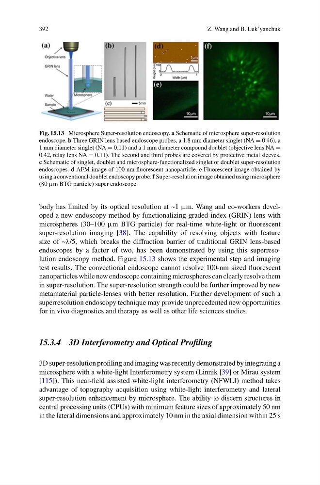
Fig. 15.13 Microsphere Super-resolution endoscopy. a Schematic of microsphere super-resolution
endoscope. b Three GRIN lens based endoscope probes, a 1.8 mm diameter singlet (NA = 0.46), a
1 mm diameter singlet (NA = 0.11) and a 1 mm diameter compound doublet (objective lens NA =
0.42, relay lens NA = 0.11). The second and third probes are covered by protective metal sleeves.
c Schematic of singlet, doublet and microsphere-functionalized singlet or doublet super-resolution
endoscopes. d AFM image of 100 nm fluorescent nanoparticle. e Fluorescent image obtained by
using a conventional doublet endoscopy probe. f Super-resolution image obtained using microsphere
(80 μm BTG particle) super endoscope
body has limited by its optical resolution at ~1 μm. Wang and co-workers developed a new endoscopy method by functionalizing graded-index (GRIN) lens with
microspheres (30–100 μm BTG particle) for real-time white-light or fluorescent
super-resolution imaging [38]. The capability of resolving objects with feature
size of ~λ/5, which breaks the diffraction barrier of traditional GRIN lens-based
endoscopes by a factor of two, has been demonstrated by using this superresolution endoscopy method. Figure 15.13 shows the experimental step and imaging
test results. The convectional endoscope cannot resolve 100-nm sized fluorescent
nanoparticles while new endoscope containing microspheres can clearly resolve them
in super-resolution. The super-resolution strength could be further improved by new
metamaterial particle-lenses with better resolution. Further development of such a
superresolution endoscopy technique may provide unprecedented new opportunities
for in vivo diagnostics and therapy as well as other life sciences studies.
15.3.4 3D Interferometry and Optical Profiling
3D super-resolution profiling and imaging was recently demonstrated by integrating a
microsphere with a white-light Interferometry system (Linnik [39] or Mirau system
[115]). This near-field assisted white-light interferometry (NFWLI) method takes
advantage of topography acquisition using white-light interferometry and lateral
super-resolution enhancement by microsphere. The ability to discern structures in
central processing units (CPUs) with minimum feature sizes of approximately 50 nm
in the lateral dimensions and approximately 10 nm in the axial dimension within 25 s
Z. Wang and B. Luk’yanchuk
Fig. 15.13 Microsphere Super-resolution endoscopy. a Schematic of microsphere super-resolution
endoscope. b Three GRIN lens based endoscope probes, a 1.8 mm diameter singlet (NA = 0.46), a
1 mm diameter singlet (NA = 0.11) and a 1 mm diameter compound doublet (objective lens NA =
0.42, relay lens NA = 0.11). The second and third probes are covered by protective metal sleeves.
c Schematic of singlet, doublet and microsphere-functionalized singlet or doublet super-resolution
endoscopes. d AFM image of 100 nm fluorescent nanoparticle. e Fluorescent image obtained by
using a conventional doublet endoscopy probe. f Super-resolution image obtained using microsphere
(80 μm BTG particle) super endoscope
body has limited by its optical resolution at ~1 μm. Wang and co-workers developed a new endoscopy method by functionalizing graded-index (GRIN) lens with
microspheres (30–100 μm BTG particle) for real-time white-light or fluorescent
super-resolution imaging [38]. The capability of resolving objects with feature
size of ~λ/5, which breaks the diffraction barrier of traditional GRIN lens-based
endoscopes by a factor of two, has been demonstrated by using this superresolution endoscopy method. Figure 15.13 shows the experimental step and imaging
test results. The convectional endoscope cannot resolve 100-nm sized fluorescent
nanoparticles while new endoscope containing microspheres can clearly resolve them
in super-resolution. The super-resolution strength could be further improved by new
metamaterial particle-lenses with better resolution. Further development of such a
superresolution endoscopy technique may provide unprecedented new opportunities
for in vivo diagnostics and therapy as well as other life sciences studies.
15.3.4 3D Interferometry and Optical Profiling
3D super-resolution profiling and imaging was recently demonstrated by integrating a
microsphere with a white-light Interferometry system (Linnik [39] or Mirau system
[115]). This near-field assisted white-light interferometry (NFWLI) method takes
advantage of topography acquisition using white-light interferometry and lateral
super-resolution enhancement by microsphere. The ability to discern structures in
central processing units (CPUs) with minimum feature sizes of approximately 50 nm
in the lateral dimensions and approximately 10 nm in the axial dimension within 25 s