

15 Super-Resolution Imaging and Microscopy by Dielectric …
391
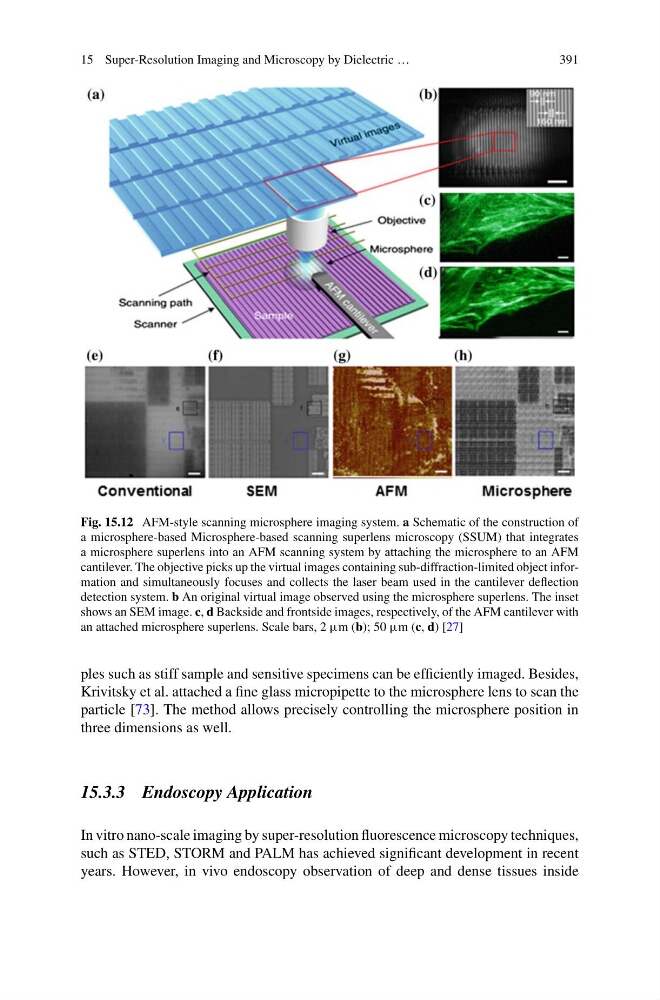
Fig. 15.12 AFM-style scanning microsphere imaging system. a Schematic of the construction of
a microsphere-based Microsphere-based scanning superlens microscopy (SSUM) that integrates
a microsphere superlens into an AFM scanning system by attaching the microsphere to an AFM
cantilever. The objective picks up the virtual images containing sub-diffraction-limited object information and simultaneously focuses and collects the laser beam used in the cantilever deflection
detection system. b An original virtual image observed using the microsphere superlens. The inset
shows an SEM image. c, d Backside and frontside images, respectively, of the AFM cantilever with
an attached microsphere superlens. Scale bars, 2 μm (b); 50 μm (c, d) [27]
ples such as stiff sample and sensitive specimens can be efficiently imaged. Besides,
Krivitsky et al. attached a fine glass micropipette to the microsphere lens to scan the
particle [73]. The method allows precisely controlling the microsphere position in
three dimensions as well.
15.3.3 Endoscopy Application
In vitro nano-scale imaging by super-resolution fluorescence microscopy techniques,
such as STED, STORM and PALM has achieved significant development in recent
years. However, in vivo endoscopy observation of deep and dense tissues inside
391
Fig. 15.12 AFM-style scanning microsphere imaging system. a Schematic of the construction of
a microsphere-based Microsphere-based scanning superlens microscopy (SSUM) that integrates
a microsphere superlens into an AFM scanning system by attaching the microsphere to an AFM
cantilever. The objective picks up the virtual images containing sub-diffraction-limited object information and simultaneously focuses and collects the laser beam used in the cantilever deflection
detection system. b An original virtual image observed using the microsphere superlens. The inset
shows an SEM image. c, d Backside and frontside images, respectively, of the AFM cantilever with
an attached microsphere superlens. Scale bars, 2 μm (b); 50 μm (c, d) [27]
ples such as stiff sample and sensitive specimens can be efficiently imaged. Besides,
Krivitsky et al. attached a fine glass micropipette to the microsphere lens to scan the
particle [73]. The method allows precisely controlling the microsphere position in
three dimensions as well.
15.3.3 Endoscopy Application
In vitro nano-scale imaging by super-resolution fluorescence microscopy techniques,
such as STED, STORM and PALM has achieved significant development in recent
years. However, in vivo endoscopy observation of deep and dense tissues inside