

14 Label-Free Super-Resolution Imaging with Hyperbolic Materials
365
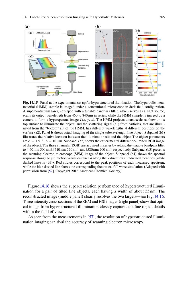
Fig. 14.15 Panel a: the experimental set up for hyperstructured illumination. The hyperbolic metamaterial (HMM) sample is imaged under a conventional microscope in dark-field configuration.
A supercontinuum laser, equipped with a tunable bandpass filter, which serves as a light source,
scans its output wavelength from 460 to 840 nm in series, while the HMM sample is imaged by a
camera to form a hyperspectral image I (x, y, λ). The HMM projects a nanoscale rainbow on its
top surface to illuminate the object, and the scattering signal (a1) from particles, that are illuminated from the “bottom” slit of the HMM, has different wavelengths at different positions on the
surface (a2). Panel b shows actual imaging of the single subwavelength line object. Subpanel (b1)
illustrates the relative location between the illumination slit and the object The object parameters
are α = 1.51 ◦ , L = 10 µm. Subpanel (b2) shows the experimental diffraction-limited RGB image
of the object. The three channels (RGB) are acquired in series by setting the tunable bandpass filter
to [460 nm: 500 nm], [510 nm: 570 nm], and [580 nm: 700 nm], respectively. Subpanel (b3) presents
the scanning electron microscope (SEM) image of the object. Subpanel (b4) shows the spectral
response along the y direction versus distance d along the x direction at indicated locations (white
dashed lines in (b3)). Red circles correspond to the peak positions of each measured spectrum,
while the blue dashed line shows the corresponding theoretical full wave simulation (Adapted with
permission from [57], Copyright 2018 American Chemical Society)
Figure 14.16 shows the super-resolution performance of hyperstructured illumination for a pair of tilted line objects, each having a width of about 35 nm. The
reconstructed image (middle panel) clearly resolves the two targets—see Fig. 14.16.
Three intensity cross sections of the SEM and HSI images (right panel) show that optical image from hyperstructured illumination closely captures the fine object details
within the field of view.
As seen from the measurements in [57], the resolution of hyperstructured illumination imaging can rival the accuracy of scanning electron microscopy.
365
Fig. 14.15 Panel a: the experimental set up for hyperstructured illumination. The hyperbolic metamaterial (HMM) sample is imaged under a conventional microscope in dark-field configuration.
A supercontinuum laser, equipped with a tunable bandpass filter, which serves as a light source,
scans its output wavelength from 460 to 840 nm in series, while the HMM sample is imaged by a
camera to form a hyperspectral image I (x, y, λ). The HMM projects a nanoscale rainbow on its
top surface to illuminate the object, and the scattering signal (a1) from particles, that are illuminated from the “bottom” slit of the HMM, has different wavelengths at different positions on the
surface (a2). Panel b shows actual imaging of the single subwavelength line object. Subpanel (b1)
illustrates the relative location between the illumination slit and the object The object parameters
are α = 1.51 ◦ , L = 10 µm. Subpanel (b2) shows the experimental diffraction-limited RGB image
of the object. The three channels (RGB) are acquired in series by setting the tunable bandpass filter
to [460 nm: 500 nm], [510 nm: 570 nm], and [580 nm: 700 nm], respectively. Subpanel (b3) presents
the scanning electron microscope (SEM) image of the object. Subpanel (b4) shows the spectral
response along the y direction versus distance d along the x direction at indicated locations (white
dashed lines in (b3)). Red circles correspond to the peak positions of each measured spectrum,
while the blue dashed line shows the corresponding theoretical full wave simulation (Adapted with
permission from [57], Copyright 2018 American Chemical Society)
Figure 14.16 shows the super-resolution performance of hyperstructured illumination for a pair of tilted line objects, each having a width of about 35 nm. The
reconstructed image (middle panel) clearly resolves the two targets—see Fig. 14.16.
Three intensity cross sections of the SEM and HSI images (right panel) show that optical image from hyperstructured illumination closely captures the fine object details
within the field of view.
As seen from the measurements in [57], the resolution of hyperstructured illumination imaging can rival the accuracy of scanning electron microscopy.