

360
E. Khan and E. Narimanov
illuminate the top surface. Since the spatial spectra of the beams are dominated by
high wavenumber components, the beamwidth, as well as the illumination spot size,
are subwavelength.
Due to the presence of loss, hyperbolic medium has strong material dispersion
(ω) owing to Kramers–Kronig relations [10]. As a result, a change in frequency will
lead to variation of the emission angle of the beams θ c (ω), as illustrated in Fig. 14.11b.
This allows one to selectively illuminate different subwavelength regions of the top
surface. Therefore by sweeping through the frequency of the illuminating light, the
entire object plane situated at the top of the hyperbolic substrate can be scanned and
a deep subwavelength image can be obtained. This hyperbolic metamaterial assisted
hyperspectral imaging process is the main idea of hyperstructured illumination [29].
Note that material dispersion, which is generally considered an engineering limitation
for many optical systems designed for a particular resonant condition, here forms a
key element to the imaging process.
Although a finite unit cell size of the metamaterial Λ will add a quantitative
correction to the dispersion, the topology of the iso-frequency surface (see Fig. 14.2b)
along with its wavelength dependence is essentially preserved, and therefore, the
beam pattern of hyperstructured illumination retains its subwavelength structure that
evolves with the wavelength variation. However, since the size of the unit cell limits
the wavenumbers in the object plane to ∼1/Λ, the final resolution attainable from
this imaging process becomes on the order of Λ, which in the current fabrication
technology [56] can reach down to a few nanometers.
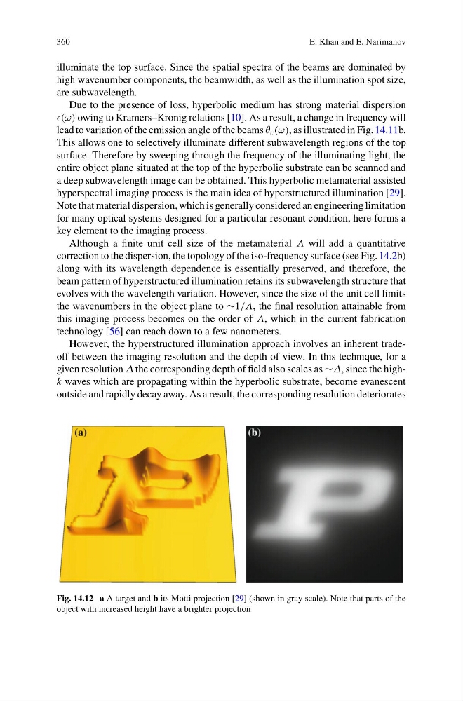
However, the hyperstructured illumination approach involves an inherent tradeoff between the imaging resolution and the depth of view. In this technique, for a
given resolution Δ the corresponding depth of field also scales as ∼Δ, since the highk waves which are propagating within the hyperbolic substrate, become evanescent
outside and rapidly decay away. As a result, the corresponding resolution deteriorates
Fig. 14.12 a A target and b its Motti projection [29] (shown in gray scale). Note that parts of the
object with increased height have a brighter projection
E. Khan and E. Narimanov
illuminate the top surface. Since the spatial spectra of the beams are dominated by
high wavenumber components, the beamwidth, as well as the illumination spot size,
are subwavelength.
Due to the presence of loss, hyperbolic medium has strong material dispersion
(ω) owing to Kramers–Kronig relations [10]. As a result, a change in frequency will
lead to variation of the emission angle of the beams θ c (ω), as illustrated in Fig. 14.11b.
This allows one to selectively illuminate different subwavelength regions of the top
surface. Therefore by sweeping through the frequency of the illuminating light, the
entire object plane situated at the top of the hyperbolic substrate can be scanned and
a deep subwavelength image can be obtained. This hyperbolic metamaterial assisted
hyperspectral imaging process is the main idea of hyperstructured illumination [29].
Note that material dispersion, which is generally considered an engineering limitation
for many optical systems designed for a particular resonant condition, here forms a
key element to the imaging process.
Although a finite unit cell size of the metamaterial Λ will add a quantitative
correction to the dispersion, the topology of the iso-frequency surface (see Fig. 14.2b)
along with its wavelength dependence is essentially preserved, and therefore, the
beam pattern of hyperstructured illumination retains its subwavelength structure that
evolves with the wavelength variation. However, since the size of the unit cell limits
the wavenumbers in the object plane to ∼1/Λ, the final resolution attainable from
this imaging process becomes on the order of Λ, which in the current fabrication
technology [56] can reach down to a few nanometers.
However, the hyperstructured illumination approach involves an inherent tradeoff between the imaging resolution and the depth of view. In this technique, for a
given resolution Δ the corresponding depth of field also scales as ∼Δ, since the highk waves which are propagating within the hyperbolic substrate, become evanescent
outside and rapidly decay away. As a result, the corresponding resolution deteriorates
Fig. 14.12 a A target and b its Motti projection [29] (shown in gray scale). Note that parts of the
object with increased height have a brighter projection