

358
E. Khan and E. Narimanov
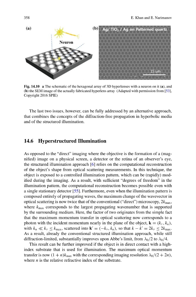
Fig. 14.10 a The schematic of the hexagonal array of 3D hyperlenses with a neuron on it (a), and
(b) the SEM image of the actually fabricated hyperlens array (Adapted with permission from [53],
Copyright 2016 SPIE)
The last two issues, however, can be fully addressed by an alternative approach,
that combines the concepts of the diffraction-free propagation in hyperbolic media
and of the structured illumination.
14.6 Hyperstructured Illumination
As opposed to the “direct” imaging where the objective is the formation of a (magnified) image on a physical screen, a detector or the retina of an observer’s eye,
the structured illumination approach [6] relies on the computational reconstruction
of the object’s shape from optical scattering measurements. In this technique, the
object is exposed to a controlled illumination pattern, which can be (rapidly) modified during the imaging. As a result, with sufficient “degrees of freedom” in the
illumination pattern, the computational reconstruction becomes possible even with
a single stationary detector [55]. Furthermore, even when the illumination pattern is
composed entirely of propagating waves, the maximum change of the wavevector in
optical scattering is now twice that of the conventional (“direct”) microscopy, 2k max ,
where k max corresponds to the largest propagating wavenumber that is supported
by the surrounding medium. Here, the factor of two originates from the simple fact
that the maximum momentum transfer in optical scattering now corresponds to a
photon with the incident momentum nearly in the plane of the object, k = (k τ , k n ),
with k n k τ ≤ k max , scattered into k
= (−k τ , k n ), so that k − k
= 2k τ ≤ 2k max .
As a result, already the conventional structured illumination approach, while still
diffraction-limited, substantially improves upon Abbe’s limit, from λ 0 /2 to λ 0 /4.
This result can be further improved if the object is in direct contact with a highindex substrate that is used for illumination. The maximum optical momentum
transfer is now (1 + n)k max with the corresponding imaging resolution λ 0 /(2 + 2n),
where n is the relative refractive index of the substrate.
E. Khan and E. Narimanov
Fig. 14.10 a The schematic of the hexagonal array of 3D hyperlenses with a neuron on it (a), and
(b) the SEM image of the actually fabricated hyperlens array (Adapted with permission from [53],
Copyright 2016 SPIE)
The last two issues, however, can be fully addressed by an alternative approach,
that combines the concepts of the diffraction-free propagation in hyperbolic media
and of the structured illumination.
14.6 Hyperstructured Illumination
As opposed to the “direct” imaging where the objective is the formation of a (magnified) image on a physical screen, a detector or the retina of an observer’s eye,
the structured illumination approach [6] relies on the computational reconstruction
of the object’s shape from optical scattering measurements. In this technique, the
object is exposed to a controlled illumination pattern, which can be (rapidly) modified during the imaging. As a result, with sufficient “degrees of freedom” in the
illumination pattern, the computational reconstruction becomes possible even with
a single stationary detector [55]. Furthermore, even when the illumination pattern is
composed entirely of propagating waves, the maximum change of the wavevector in
optical scattering is now twice that of the conventional (“direct”) microscopy, 2k max ,
where k max corresponds to the largest propagating wavenumber that is supported
by the surrounding medium. Here, the factor of two originates from the simple fact
that the maximum momentum transfer in optical scattering now corresponds to a
photon with the incident momentum nearly in the plane of the object, k = (k τ , k n ),
with k n k τ ≤ k max , scattered into k
= (−k τ , k n ), so that k − k
= 2k τ ≤ 2k max .
As a result, already the conventional structured illumination approach, while still
diffraction-limited, substantially improves upon Abbe’s limit, from λ 0 /2 to λ 0 /4.
This result can be further improved if the object is in direct contact with a highindex substrate that is used for illumination. The maximum optical momentum
transfer is now (1 + n)k max with the corresponding imaging resolution λ 0 /(2 + 2n),
where n is the relative refractive index of the substrate.