

13 Super-Resolution Microscopy Techniques Based …
333
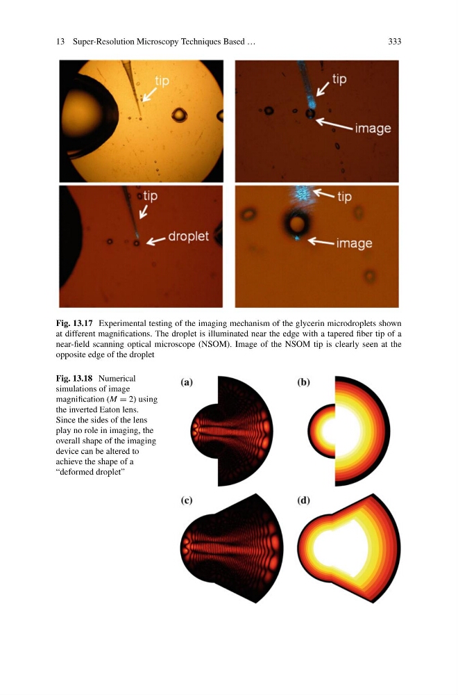
Fig. 13.17 Experimental testing of the imaging mechanism of the glycerin microdroplets shown
at different magnifications. The droplet is illuminated near the edge with a tapered fiber tip of a
near-field scanning optical microscope (NSOM). Image of the NSOM tip is clearly seen at the
opposite edge of the droplet
Fig. 13.18 Numerical
simulations of image
magnification (M = 2) using
the inverted Eaton lens.
Since the sides of the lens
play no role in imaging, the
overall shape of the imaging
device can be altered to
achieve the shape of a
“deformed droplet”
333
Fig. 13.17 Experimental testing of the imaging mechanism of the glycerin microdroplets shown
at different magnifications. The droplet is illuminated near the edge with a tapered fiber tip of a
near-field scanning optical microscope (NSOM). Image of the NSOM tip is clearly seen at the
opposite edge of the droplet
Fig. 13.18 Numerical
simulations of image
magnification (M = 2) using
the inverted Eaton lens.
Since the sides of the lens
play no role in imaging, the
overall shape of the imaging
device can be altered to
achieve the shape of a
“deformed droplet”