

322
I. I. Smolyaninov and V. N. Smolyaninova
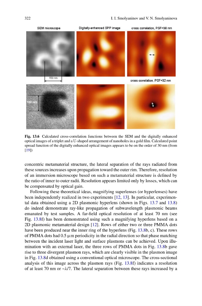
Fig. 13.6 Calculated cross-correlation functions between the SEM and the digitally enhanced
optical images of a triplet and a U-shaped arrangement of nanoholes in a gold film. Calculated point
spread function of the digitally enhanced optical images appears to be on the order of 30 nm (from
[19])
concentric metamaterial structure, the lateral separation of the rays radiated from
these sources increases upon propagation toward the outer rim. Therefore, resolution
of an immersion microscope based on such a metamaterial structure is defined by
the ratio of inner to outer radii. Resolution appears limited only by losses, which can
be compensated by optical gain.
Following these theoretical ideas, magnifying superlenses (or hyperlenses) have
been independently realized in two experiments [12, 13]. In particular, experimental data obtained using a 2D plasmonic hyperlens (shown in Figs. 13.7 and 13.8)
do indeed demonstrate ray-like propagation of subwavelength plasmonic beams
emanated by test samples. A far-field optical resolution of at least 70 nm (see
Fig. 13.8f) has been demonstrated using such a magnifying hyperlens based on a
2D plasmonic metamaterial design [12]. Rows of either two or three PMMA dots
have been produced near the inner ring of the hyperlens (Fig. 13.8b, c). These rows
of PMMA dots had 0.5 µm periodicity in the radial direction so that phase matching
between the incident laser light and surface plasmons can be achieved. Upon illumination with an external laser, the three rows of PMMA dots in Fig. 13.8b gave
rise to three divergent plasmon rays, which are clearly visible in the plasmon image
in Fig. 13.8d obtained using a conventional optical microscope. The cross-sectional
analysis of this image across the plasmon rays (Fig. 13.8f) indicates a resolution
of at least 70 nm or ~λ/7. The lateral separation between these rays increased by a
I. I. Smolyaninov and V. N. Smolyaninova
Fig. 13.6 Calculated cross-correlation functions between the SEM and the digitally enhanced
optical images of a triplet and a U-shaped arrangement of nanoholes in a gold film. Calculated point
spread function of the digitally enhanced optical images appears to be on the order of 30 nm (from
[19])
concentric metamaterial structure, the lateral separation of the rays radiated from
these sources increases upon propagation toward the outer rim. Therefore, resolution
of an immersion microscope based on such a metamaterial structure is defined by
the ratio of inner to outer radii. Resolution appears limited only by losses, which can
be compensated by optical gain.
Following these theoretical ideas, magnifying superlenses (or hyperlenses) have
been independently realized in two experiments [12, 13]. In particular, experimental data obtained using a 2D plasmonic hyperlens (shown in Figs. 13.7 and 13.8)
do indeed demonstrate ray-like propagation of subwavelength plasmonic beams
emanated by test samples. A far-field optical resolution of at least 70 nm (see
Fig. 13.8f) has been demonstrated using such a magnifying hyperlens based on a
2D plasmonic metamaterial design [12]. Rows of either two or three PMMA dots
have been produced near the inner ring of the hyperlens (Fig. 13.8b, c). These rows
of PMMA dots had 0.5 µm periodicity in the radial direction so that phase matching
between the incident laser light and surface plasmons can be achieved. Upon illumination with an external laser, the three rows of PMMA dots in Fig. 13.8b gave
rise to three divergent plasmon rays, which are clearly visible in the plasmon image
in Fig. 13.8d obtained using a conventional optical microscope. The cross-sectional
analysis of this image across the plasmon rays (Fig. 13.8f) indicates a resolution
of at least 70 nm or ~λ/7. The lateral separation between these rays increased by a