

13 Super-Resolution Microscopy Techniques Based …
321
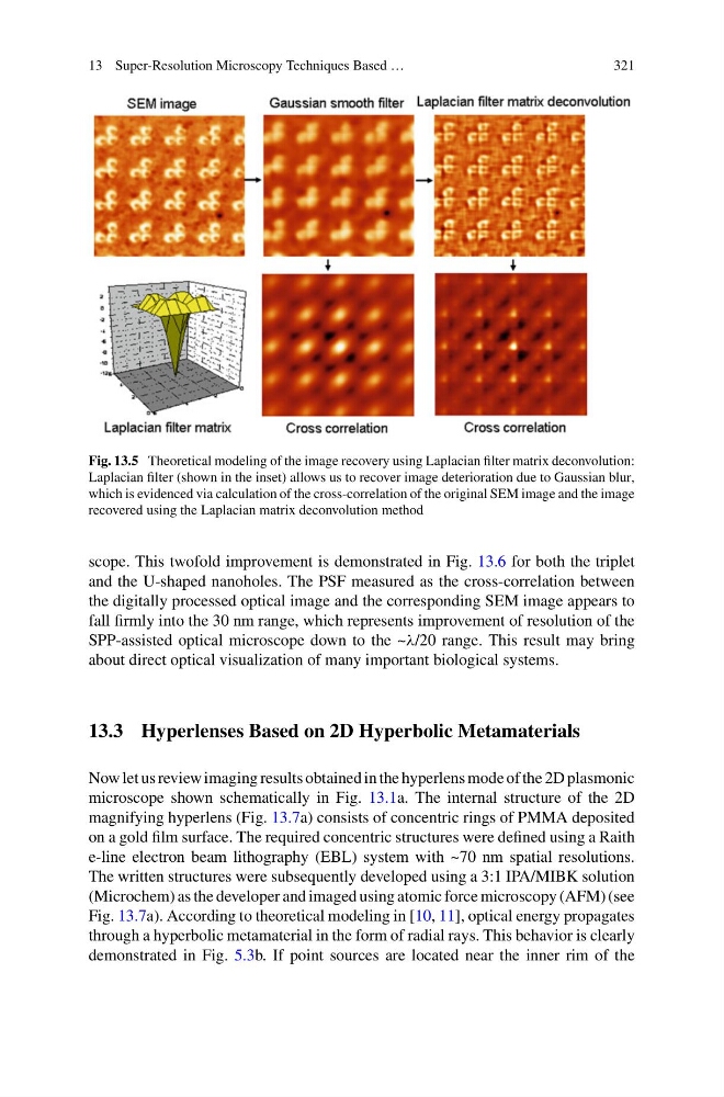
Fig. 13.5 Theoretical modeling of the image recovery using Laplacian filter matrix deconvolution:
Laplacian filter (shown in the inset) allows us to recover image deterioration due to Gaussian blur,
which is evidenced via calculation of the cross-correlation of the original SEM image and the image
recovered using the Laplacian matrix deconvolution method
scope. This twofold improvement is demonstrated in Fig. 13.6 for both the triplet
and the U-shaped nanoholes. The PSF measured as the cross-correlation between
the digitally processed optical image and the corresponding SEM image appears to
fall firmly into the 30 nm range, which represents improvement of resolution of the
SPP-assisted optical microscope down to the ~λ/20 range. This result may bring
about direct optical visualization of many important biological systems.
13.3 Hyperlenses Based on 2D Hyperbolic Metamaterials
Now let us review imaging results obtained in the hyperlens mode of the 2D plasmonic
microscope shown schematically in Fig. 13.1a. The internal structure of the 2D
magnifying hyperlens (Fig. 13.7a) consists of concentric rings of PMMA deposited
on a gold film surface. The required concentric structures were defined using a Raith
e-line electron beam lithography (EBL) system with ~70 nm spatial resolutions.
The written structures were subsequently developed using a 3:1 IPA/MIBK solution
(Microchem) as the developer and imaged using atomic force microscopy (AFM) (see
Fig. 13.7a). According to theoretical modeling in [10, 11], optical energy propagates
through a hyperbolic metamaterial in the form of radial rays. This behavior is clearly
demonstrated in Fig. 5.3b. If point sources are located near the inner rim of the
321
Fig. 13.5 Theoretical modeling of the image recovery using Laplacian filter matrix deconvolution:
Laplacian filter (shown in the inset) allows us to recover image deterioration due to Gaussian blur,
which is evidenced via calculation of the cross-correlation of the original SEM image and the image
recovered using the Laplacian matrix deconvolution method
scope. This twofold improvement is demonstrated in Fig. 13.6 for both the triplet
and the U-shaped nanoholes. The PSF measured as the cross-correlation between
the digitally processed optical image and the corresponding SEM image appears to
fall firmly into the 30 nm range, which represents improvement of resolution of the
SPP-assisted optical microscope down to the ~λ/20 range. This result may bring
about direct optical visualization of many important biological systems.
13.3 Hyperlenses Based on 2D Hyperbolic Metamaterials
Now let us review imaging results obtained in the hyperlens mode of the 2D plasmonic
microscope shown schematically in Fig. 13.1a. The internal structure of the 2D
magnifying hyperlens (Fig. 13.7a) consists of concentric rings of PMMA deposited
on a gold film surface. The required concentric structures were defined using a Raith
e-line electron beam lithography (EBL) system with ~70 nm spatial resolutions.
The written structures were subsequently developed using a 3:1 IPA/MIBK solution
(Microchem) as the developer and imaged using atomic force microscopy (AFM) (see
Fig. 13.7a). According to theoretical modeling in [10, 11], optical energy propagates
through a hyperbolic metamaterial in the form of radial rays. This behavior is clearly
demonstrated in Fig. 5.3b. If point sources are located near the inner rim of the