

320
I. I. Smolyaninov and V. N. Smolyaninova
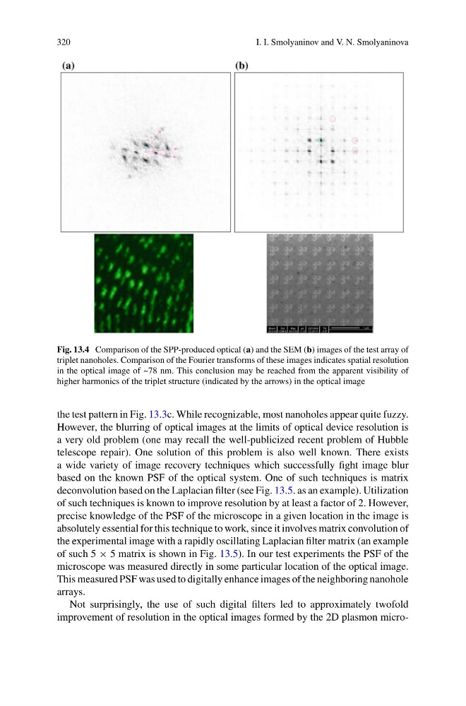
Fig. 13.4 Comparison of the SPP-produced optical (a) and the SEM (b) images of the test array of
triplet nanoholes. Comparison of the Fourier transforms of these images indicates spatial resolution
in the optical image of ~78 nm. This conclusion may be reached from the apparent visibility of
higher harmonics of the triplet structure (indicated by the arrows) in the optical image
the test pattern in Fig. 13.3c. While recognizable, most nanoholes appear quite fuzzy.
However, the blurring of optical images at the limits of optical device resolution is
a very old problem (one may recall the well-publicized recent problem of Hubble
telescope repair). One solution of this problem is also well known. There exists
a wide variety of image recovery techniques which successfully fight image blur
based on the known PSF of the optical system. One of such techniques is matrix
deconvolution based on the Laplacian filter (see Fig. 13.5. as an example). Utilization
of such techniques is known to improve resolution by at least a factor of 2. However,
precise knowledge of the PSF of the microscope in a given location in the image is
absolutely essential for this technique to work, since it involves matrix convolution of
the experimental image with a rapidly oscillating Laplacian filter matrix (an example
of such 5 × 5 matrix is shown in Fig. 13.5). In our test experiments the PSF of the
microscope was measured directly in some particular location of the optical image.
This measured PSF was used to digitally enhance images of the neighboring nanohole
arrays.
Not surprisingly, the use of such digital filters led to approximately twofold
improvement of resolution in the optical images formed by the 2D plasmon micro-
I. I. Smolyaninov and V. N. Smolyaninova
Fig. 13.4 Comparison of the SPP-produced optical (a) and the SEM (b) images of the test array of
triplet nanoholes. Comparison of the Fourier transforms of these images indicates spatial resolution
in the optical image of ~78 nm. This conclusion may be reached from the apparent visibility of
higher harmonics of the triplet structure (indicated by the arrows) in the optical image
the test pattern in Fig. 13.3c. While recognizable, most nanoholes appear quite fuzzy.
However, the blurring of optical images at the limits of optical device resolution is
a very old problem (one may recall the well-publicized recent problem of Hubble
telescope repair). One solution of this problem is also well known. There exists
a wide variety of image recovery techniques which successfully fight image blur
based on the known PSF of the optical system. One of such techniques is matrix
deconvolution based on the Laplacian filter (see Fig. 13.5. as an example). Utilization
of such techniques is known to improve resolution by at least a factor of 2. However,
precise knowledge of the PSF of the microscope in a given location in the image is
absolutely essential for this technique to work, since it involves matrix convolution of
the experimental image with a rapidly oscillating Laplacian filter matrix (an example
of such 5 × 5 matrix is shown in Fig. 13.5). In our test experiments the PSF of the
microscope was measured directly in some particular location of the optical image.
This measured PSF was used to digitally enhance images of the neighboring nanohole
arrays.
Not surprisingly, the use of such digital filters led to approximately twofold
improvement of resolution in the optical images formed by the 2D plasmon micro-