

13 Super-Resolution Microscopy Techniques Based …
319
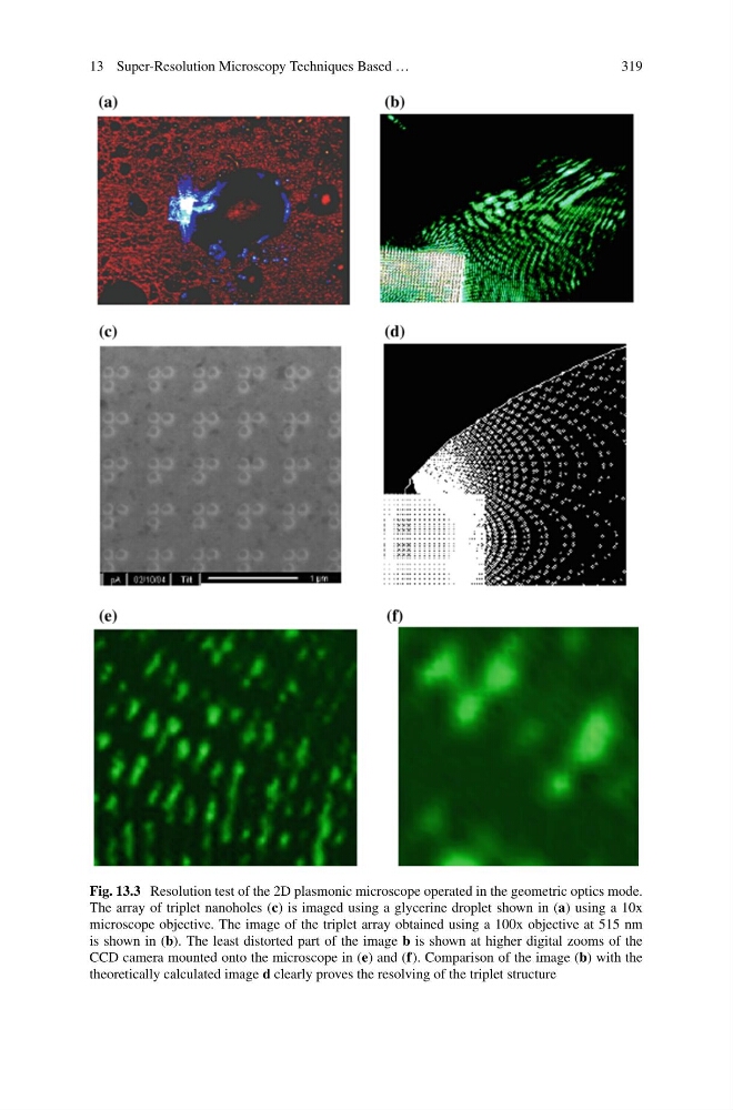
Fig. 13.3 Resolution test of the 2D plasmonic microscope operated in the geometric optics mode.
The array of triplet nanoholes (c) is imaged using a glycerine droplet shown in (a) using a 10x
microscope objective. The image of the triplet array obtained using a 100x objective at 515 nm
is shown in (b). The least distorted part of the image b is shown at higher digital zooms of the
CCD camera mounted onto the microscope in (e) and (f). Comparison of the image (b) with the
theoretically calculated image d clearly proves the resolving of the triplet structure
319
Fig. 13.3 Resolution test of the 2D plasmonic microscope operated in the geometric optics mode.
The array of triplet nanoholes (c) is imaged using a glycerine droplet shown in (a) using a 10x
microscope objective. The image of the triplet array obtained using a 100x objective at 515 nm
is shown in (b). The least distorted part of the image b is shown at higher digital zooms of the
CCD camera mounted onto the microscope in (e) and (f). Comparison of the image (b) with the
theoretically calculated image d clearly proves the resolving of the triplet structure