

1 Quantitative Phase Imaging: Principles and Applications
11
I (x, y, δ) = |U i (x, y)|
2
+ |U R |
2
+ 2|U i (x, y)||U R | cos[ϕ(x, y) + δ]
(1.17)
In general, a phase map could be easily obtained using 4 intensity measurements,
each with an increment phase delay of π /2, as
ϕ(x, y) = arg
I 0 − I π , I 3π/2 − I π/2
(1.18)
Spatial light interference microscopy (SLIM) [53] is a phase-shifting, but also
common-path QPI system. Based on an existing Zernike microscope, SLIM combines advantages of phase-contrast microscopy and Gabor’s holography to obtain
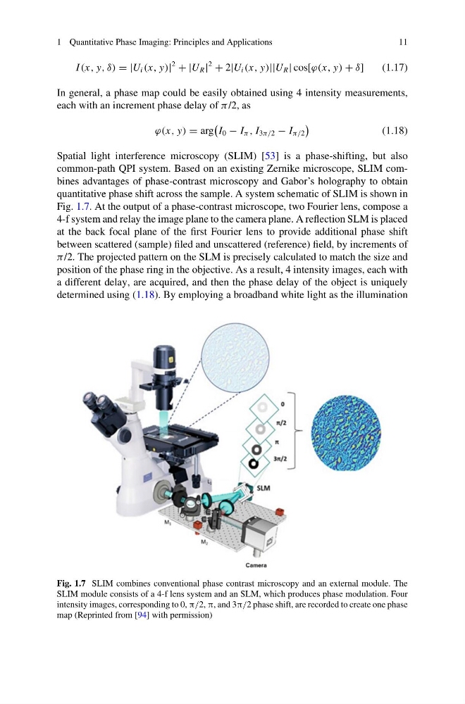
quantitative phase shift across the sample. A system schematic of SLIM is shown in
Fig. 1.7. At the output of a phase-contrast microscope, two Fourier lens, compose a
4-f system and relay the image plane to the camera plane. A reflection SLM is placed
at the back focal plane of the first Fourier lens to provide additional phase shift
between scattered (sample) filed and unscattered (reference) field, by increments of
π /2. The projected pattern on the SLM is precisely calculated to match the size and
position of the phase ring in the objective. As a result, 4 intensity images, each with
a different delay, are acquired, and then the phase delay of the object is uniquely
determined using (1.18). By employing a broadband white light as the illumination
Fig. 1.7 SLIM combines conventional phase contrast microscopy and an external module. The
SLIM module consists of a 4-f lens system and an SLM, which produces phase modulation. Four
intensity images, corresponding to 0, π/2, π, and 3π/2 phase shift, are recorded to create one phase
map (Reprinted from [94] with permission)
11
I (x, y, δ) = |U i (x, y)|
2
+ |U R |
2
+ 2|U i (x, y)||U R | cos[ϕ(x, y) + δ]
(1.17)
In general, a phase map could be easily obtained using 4 intensity measurements,
each with an increment phase delay of π /2, as
ϕ(x, y) = arg
I 0 − I π , I 3π/2 − I π/2
(1.18)
Spatial light interference microscopy (SLIM) [53] is a phase-shifting, but also
common-path QPI system. Based on an existing Zernike microscope, SLIM combines advantages of phase-contrast microscopy and Gabor’s holography to obtain
quantitative phase shift across the sample. A system schematic of SLIM is shown in
Fig. 1.7. At the output of a phase-contrast microscope, two Fourier lens, compose a
4-f system and relay the image plane to the camera plane. A reflection SLM is placed
at the back focal plane of the first Fourier lens to provide additional phase shift
between scattered (sample) filed and unscattered (reference) field, by increments of
π /2. The projected pattern on the SLM is precisely calculated to match the size and
position of the phase ring in the objective. As a result, 4 intensity images, each with
a different delay, are acquired, and then the phase delay of the object is uniquely
determined using (1.18). By employing a broadband white light as the illumination
Fig. 1.7 SLIM combines conventional phase contrast microscopy and an external module. The
SLIM module consists of a 4-f lens system and an SLM, which produces phase modulation. Four
intensity images, corresponding to 0, π/2, π, and 3π/2 phase shift, are recorded to create one phase
map (Reprinted from [94] with permission)