

1 Quantitative Phase Imaging: Principles and Applications
9
phase map, ϕ (Fig. 1.4c). Other reconstruction methods are discussed elsewhere
[28–32].
The off-axis method reconstructs a phase map from one raw measurement, and
the speed of this imaging modality is only limited by the photodetector, which makes
it widely adopted [33–40]. However, the high time-bandwidth product comes at the
expense of the space-bandwidth product, as the interferogram period must be sampled
by a sufficient number of pixels as described in [28].
Most off-axis methods use physically separated beams, specific to traditional
interferometers, e.g., Michelson and Mach–Zehnder, which results in noisy measurements. One of the successful demonstrations of common-path, off-axis QPI is
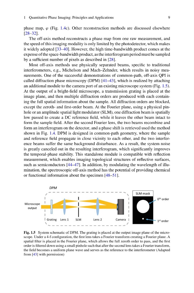
called diffraction phase microscopy (DPM) [41–43], which is realized by attaching
an additional module to the camera port of an existing microscope system (Fig. 1.5).
At the output of a bright-field microscope, a transmission grating is placed at the
image plane, and then multiple diffraction orders are produced with each containing the full spatial information about the sample. All diffraction orders are blocked,
except the zeroth- and first-order beam. At the Fourier plane, using a physical pinhole or an amplitude spatial light modulator (SLM), one diffraction beam is spatially
low passed to create a DC reference field, while it leaves the other beam intact to
form the sample field. After the second Fourier lens, the two beams recombine and
form an interferogram on the detector, and a phase shift is retrieved used the method
shown in Fig. 1.4. DPM is designed in common-path geometry, where the sample
and reference field propagate in close vicinity to each other, and the two interference beams suffer the same background disturbance. As a result, the system noise
is greatly canceled out in the resulting interferogram, which significantly improves
the temporal-phase stability. This standalone module is compatible with reflection
measurement, which enables imaging topological structures of reflective surfaces,
such as semiconductors [44–47]. In addition, by modulating the wavelength of illumination, the spectroscopic off-axis method has the potential of providing chemical
or functional information about the specimen [48–51].
Fig. 1.5 System schematic of DPM. The grating is placed at the output image plane of the microscope. Under a 4-f configuration, the first lens takes a Fourier transform creating a Fourier plane. A
spatial filter is placed in the Fourier plane, which allows the full zeroth order to pass, and the first
order is filtered down using a small pinhole such that after the second lens takes a Fourier transform,
the field becomes a uniform plane wave and serves as the reference to the interferometer (Adapted
from [43] with permission)
9
phase map, ϕ (Fig. 1.4c). Other reconstruction methods are discussed elsewhere
[28–32].
The off-axis method reconstructs a phase map from one raw measurement, and
the speed of this imaging modality is only limited by the photodetector, which makes
it widely adopted [33–40]. However, the high time-bandwidth product comes at the
expense of the space-bandwidth product, as the interferogram period must be sampled
by a sufficient number of pixels as described in [28].
Most off-axis methods use physically separated beams, specific to traditional
interferometers, e.g., Michelson and Mach–Zehnder, which results in noisy measurements. One of the successful demonstrations of common-path, off-axis QPI is
called diffraction phase microscopy (DPM) [41–43], which is realized by attaching
an additional module to the camera port of an existing microscope system (Fig. 1.5).
At the output of a bright-field microscope, a transmission grating is placed at the
image plane, and then multiple diffraction orders are produced with each containing the full spatial information about the sample. All diffraction orders are blocked,
except the zeroth- and first-order beam. At the Fourier plane, using a physical pinhole or an amplitude spatial light modulator (SLM), one diffraction beam is spatially
low passed to create a DC reference field, while it leaves the other beam intact to
form the sample field. After the second Fourier lens, the two beams recombine and
form an interferogram on the detector, and a phase shift is retrieved used the method
shown in Fig. 1.4. DPM is designed in common-path geometry, where the sample
and reference field propagate in close vicinity to each other, and the two interference beams suffer the same background disturbance. As a result, the system noise
is greatly canceled out in the resulting interferogram, which significantly improves
the temporal-phase stability. This standalone module is compatible with reflection
measurement, which enables imaging topological structures of reflective surfaces,
such as semiconductors [44–47]. In addition, by modulating the wavelength of illumination, the spectroscopic off-axis method has the potential of providing chemical
or functional information about the specimen [48–51].
Fig. 1.5 System schematic of DPM. The grating is placed at the output image plane of the microscope. Under a 4-f configuration, the first lens takes a Fourier transform creating a Fourier plane. A
spatial filter is placed in the Fourier plane, which allows the full zeroth order to pass, and the first
order is filtered down using a small pinhole such that after the second lens takes a Fourier transform,
the field becomes a uniform plane wave and serves as the reference to the interferometer (Adapted
from [43] with permission)