

218
H. Pinhas et al.
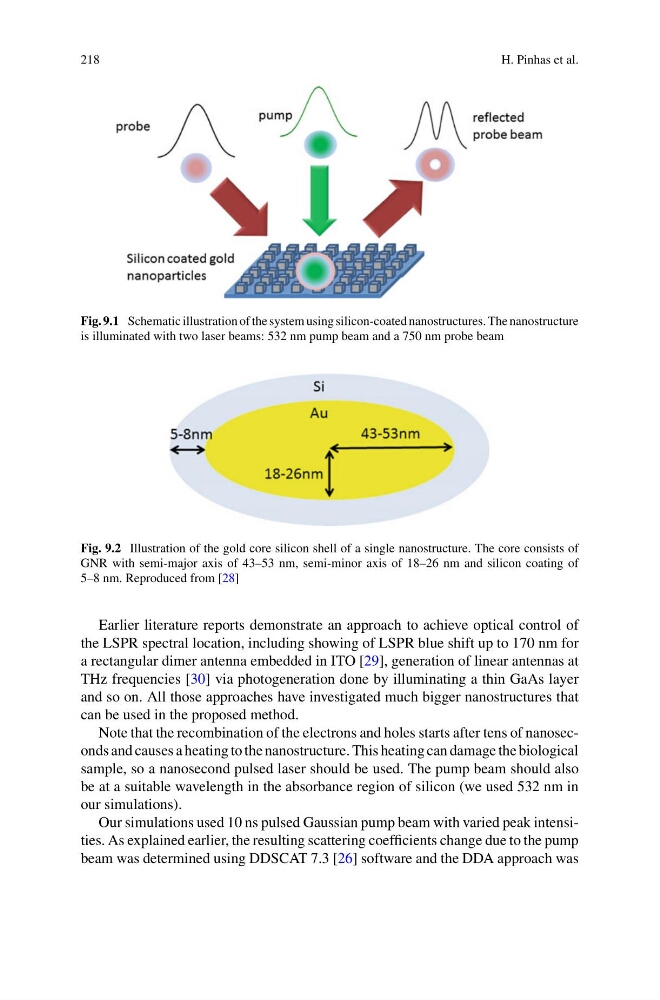
Fig. 9.1 Schematic illustration of the system using silicon-coated nanostructures. The nanostructure
is illuminated with two laser beams: 532 nm pump beam and a 750 nm probe beam
Fig. 9.2 Illustration of the gold core silicon shell of a single nanostructure. The core consists of
GNR with semi-major axis of 43–53 nm, semi-minor axis of 18–26 nm and silicon coating of
5–8 nm. Reproduced from [28]
Earlier literature reports demonstrate an approach to achieve optical control of
the LSPR spectral location, including showing of LSPR blue shift up to 170 nm for
a rectangular dimer antenna embedded in ITO [29], generation of linear antennas at
THz frequencies [30] via photogeneration done by illuminating a thin GaAs layer
and so on. All those approaches have investigated much bigger nanostructures that
can be used in the proposed method.
Note that the recombination of the electrons and holes starts after tens of nanoseconds and causes a heating to the nanostructure. This heating can damage the biological
sample, so a nanosecond pulsed laser should be used. The pump beam should also
be at a suitable wavelength in the absorbance region of silicon (we used 532 nm in
our simulations).
Our simulations used 10 ns pulsed Gaussian pump beam with varied peak intensities. As explained earlier, the resulting scattering coefficients change due to the pump
beam was determined using DDSCAT 7.3 [26] software and the DDA approach was
H. Pinhas et al.
Fig. 9.1 Schematic illustration of the system using silicon-coated nanostructures. The nanostructure
is illuminated with two laser beams: 532 nm pump beam and a 750 nm probe beam
Fig. 9.2 Illustration of the gold core silicon shell of a single nanostructure. The core consists of
GNR with semi-major axis of 43–53 nm, semi-minor axis of 18–26 nm and silicon coating of
5–8 nm. Reproduced from [28]
Earlier literature reports demonstrate an approach to achieve optical control of
the LSPR spectral location, including showing of LSPR blue shift up to 170 nm for
a rectangular dimer antenna embedded in ITO [29], generation of linear antennas at
THz frequencies [30] via photogeneration done by illuminating a thin GaAs layer
and so on. All those approaches have investigated much bigger nanostructures that
can be used in the proposed method.
Note that the recombination of the electrons and holes starts after tens of nanoseconds and causes a heating to the nanostructure. This heating can damage the biological
sample, so a nanosecond pulsed laser should be used. The pump beam should also
be at a suitable wavelength in the absorbance region of silicon (we used 532 nm in
our simulations).
Our simulations used 10 ns pulsed Gaussian pump beam with varied peak intensities. As explained earlier, the resulting scattering coefficients change due to the pump
beam was determined using DDSCAT 7.3 [26] software and the DDA approach was