

8 Super-Resolution Imaging in Raman Microscopy
207
8.4 Localization Microscopy
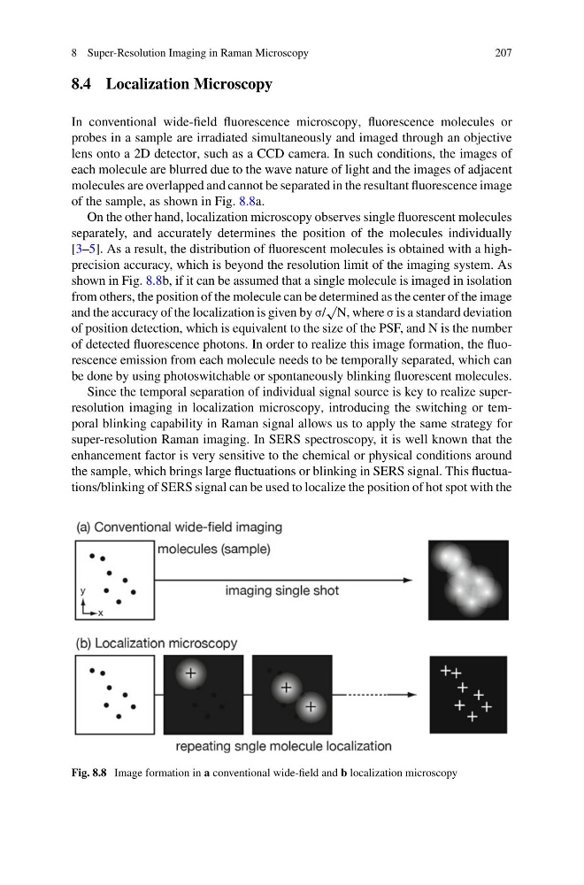
In conventional wide-field fluorescence microscopy, fluorescence molecules or
probes in a sample are irradiated simultaneously and imaged through an objective
lens onto a 2D detector, such as a CCD camera. In such conditions, the images of
each molecule are blurred due to the wave nature of light and the images of adjacent
molecules are overlapped and cannot be separated in the resultant fluorescence image
of the sample, as shown in Fig. 8.8a.
On the other hand, localization microscopy observes single fluorescent molecules
separately, and accurately determines the position of the molecules individually
[3–5]. As a result, the distribution of fluorescent molecules is obtained with a highprecision accuracy, which is beyond the resolution limit of the imaging system. As
shown in Fig. 8.8b, if it can be assumed that a single molecule is imaged in isolation
from others, the position of the molecule can be determined as the center of the image
and the accuracy of the localization is given by σ/
√
N, where σ is a standard deviation
of position detection, which is equivalent to the size of the PSF, and N is the number
of detected fluorescence photons. In order to realize this image formation, the fluorescence emission from each molecule needs to be temporally separated, which can
be done by using photoswitchable or spontaneously blinking fluorescent molecules.
Since the temporal separation of individual signal source is key to realize superresolution imaging in localization microscopy, introducing the switching or temporal blinking capability in Raman signal allows us to apply the same strategy for
super-resolution Raman imaging. In SERS spectroscopy, it is well known that the
enhancement factor is very sensitive to the chemical or physical conditions around
the sample, which brings large fluctuations or blinking in SERS signal. This fluctuations/blinking of SERS signal can be used to localize the position of hot spot with the
Fig. 8.8 Image formation in a conventional wide-field and b localization microscopy
207
8.4 Localization Microscopy
In conventional wide-field fluorescence microscopy, fluorescence molecules or
probes in a sample are irradiated simultaneously and imaged through an objective
lens onto a 2D detector, such as a CCD camera. In such conditions, the images of
each molecule are blurred due to the wave nature of light and the images of adjacent
molecules are overlapped and cannot be separated in the resultant fluorescence image
of the sample, as shown in Fig. 8.8a.
On the other hand, localization microscopy observes single fluorescent molecules
separately, and accurately determines the position of the molecules individually
[3–5]. As a result, the distribution of fluorescent molecules is obtained with a highprecision accuracy, which is beyond the resolution limit of the imaging system. As
shown in Fig. 8.8b, if it can be assumed that a single molecule is imaged in isolation
from others, the position of the molecule can be determined as the center of the image
and the accuracy of the localization is given by σ/
√
N, where σ is a standard deviation
of position detection, which is equivalent to the size of the PSF, and N is the number
of detected fluorescence photons. In order to realize this image formation, the fluorescence emission from each molecule needs to be temporally separated, which can
be done by using photoswitchable or spontaneously blinking fluorescent molecules.
Since the temporal separation of individual signal source is key to realize superresolution imaging in localization microscopy, introducing the switching or temporal blinking capability in Raman signal allows us to apply the same strategy for
super-resolution Raman imaging. In SERS spectroscopy, it is well known that the
enhancement factor is very sensitive to the chemical or physical conditions around
the sample, which brings large fluctuations or blinking in SERS signal. This fluctuations/blinking of SERS signal can be used to localize the position of hot spot with the
Fig. 8.8 Image formation in a conventional wide-field and b localization microscopy