

8 Super-Resolution Imaging in Raman Microscopy
201
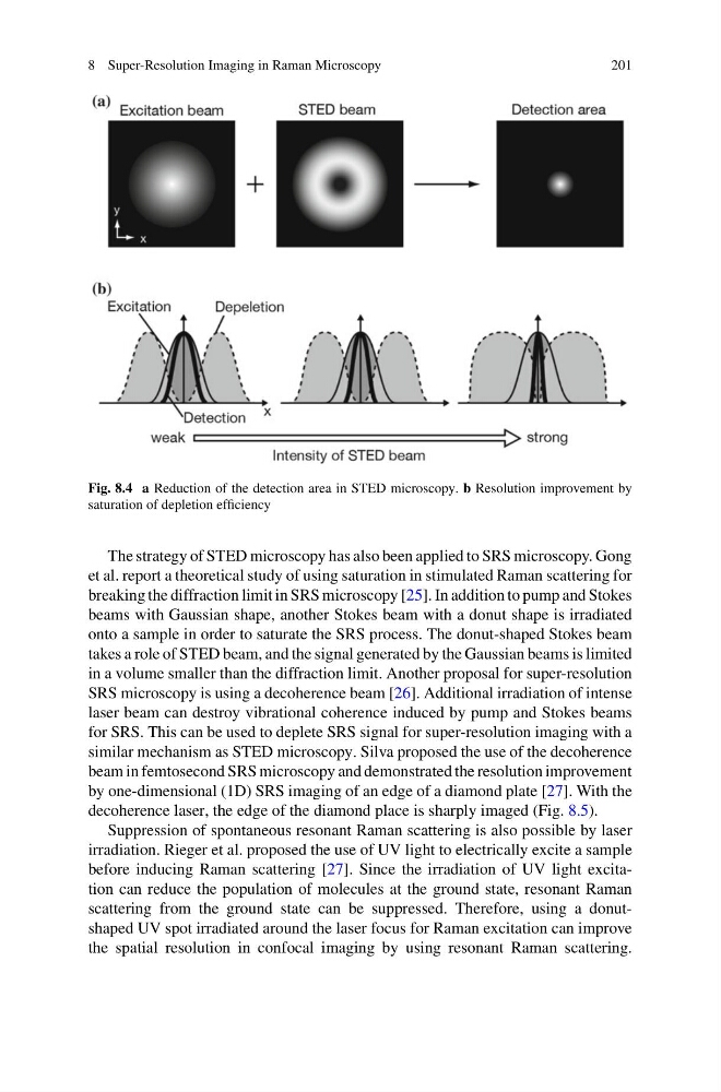
Fig. 8.4 a Reduction of the detection area in STED microscopy. b Resolution improvement by
saturation of depletion efficiency
The strategy of STED microscopy has also been applied to SRS microscopy. Gong
et al. report a theoretical study of using saturation in stimulated Raman scattering for
breaking the diffraction limit in SRS microscopy [25]. In addition to pump and Stokes
beams with Gaussian shape, another Stokes beam with a donut shape is irradiated
onto a sample in order to saturate the SRS process. The donut-shaped Stokes beam
takes a role of STED beam, and the signal generated by the Gaussian beams is limited
in a volume smaller than the diffraction limit. Another proposal for super-resolution
SRS microscopy is using a decoherence beam [26]. Additional irradiation of intense
laser beam can destroy vibrational coherence induced by pump and Stokes beams
for SRS. This can be used to deplete SRS signal for super-resolution imaging with a
similar mechanism as STED microscopy. Silva proposed the use of the decoherence
beam in femtosecond SRS microscopy and demonstrated the resolution improvement
by one-dimensional (1D) SRS imaging of an edge of a diamond plate [27]. With the
decoherence laser, the edge of the diamond place is sharply imaged (Fig. 8.5).
Suppression of spontaneous resonant Raman scattering is also possible by laser
irradiation. Rieger et al. proposed the use of UV light to electrically excite a sample
before inducing Raman scattering [27]. Since the irradiation of UV light excitation can reduce the population of molecules at the ground state, resonant Raman
scattering from the ground state can be suppressed. Therefore, using a donutshaped UV spot irradiated around the laser focus for Raman excitation can improve
the spatial resolution in confocal imaging by using resonant Raman scattering.
201
Fig. 8.4 a Reduction of the detection area in STED microscopy. b Resolution improvement by
saturation of depletion efficiency
The strategy of STED microscopy has also been applied to SRS microscopy. Gong
et al. report a theoretical study of using saturation in stimulated Raman scattering for
breaking the diffraction limit in SRS microscopy [25]. In addition to pump and Stokes
beams with Gaussian shape, another Stokes beam with a donut shape is irradiated
onto a sample in order to saturate the SRS process. The donut-shaped Stokes beam
takes a role of STED beam, and the signal generated by the Gaussian beams is limited
in a volume smaller than the diffraction limit. Another proposal for super-resolution
SRS microscopy is using a decoherence beam [26]. Additional irradiation of intense
laser beam can destroy vibrational coherence induced by pump and Stokes beams
for SRS. This can be used to deplete SRS signal for super-resolution imaging with a
similar mechanism as STED microscopy. Silva proposed the use of the decoherence
beam in femtosecond SRS microscopy and demonstrated the resolution improvement
by one-dimensional (1D) SRS imaging of an edge of a diamond plate [27]. With the
decoherence laser, the edge of the diamond place is sharply imaged (Fig. 8.5).
Suppression of spontaneous resonant Raman scattering is also possible by laser
irradiation. Rieger et al. proposed the use of UV light to electrically excite a sample
before inducing Raman scattering [27]. Since the irradiation of UV light excitation can reduce the population of molecules at the ground state, resonant Raman
scattering from the ground state can be suppressed. Therefore, using a donutshaped UV spot irradiated around the laser focus for Raman excitation can improve
the spatial resolution in confocal imaging by using resonant Raman scattering.