

156
C. Li and J.-X. Cheng
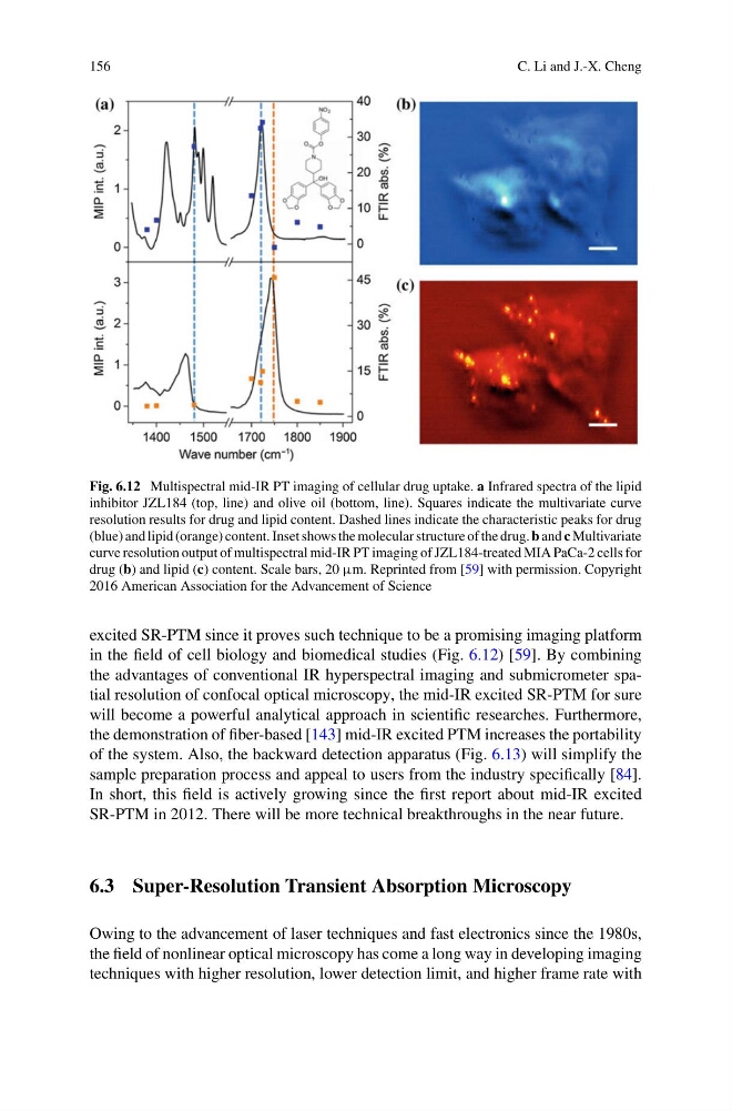
Fig. 6.12 Multispectral mid-IR PT imaging of cellular drug uptake. a Infrared spectra of the lipid
inhibitor JZL184 (top, line) and olive oil (bottom, line). Squares indicate the multivariate curve
resolution results for drug and lipid content. Dashed lines indicate the characteristic peaks for drug
(blue) and lipid (orange) content. Inset shows the molecular structure of the drug. b and c Multivariate
curve resolution output of multispectral mid-IR PT imaging of JZL184-treated MIA PaCa-2 cells for
drug (b) and lipid (c) content. Scale bars, 20 µm. Reprinted from [59] with permission. Copyright
2016 American Association for the Advancement of Science
excited SR-PTM since it proves such technique to be a promising imaging platform
in the field of cell biology and biomedical studies (Fig. 6.12) [59]. By combining
the advantages of conventional IR hyperspectral imaging and submicrometer spatial resolution of confocal optical microscopy, the mid-IR excited SR-PTM for sure
will become a powerful analytical approach in scientific researches. Furthermore,
the demonstration of fiber-based [143] mid-IR excited PTM increases the portability
of the system. Also, the backward detection apparatus (Fig. 6.13) will simplify the
sample preparation process and appeal to users from the industry specifically [84].
In short, this field is actively growing since the first report about mid-IR excited
SR-PTM in 2012. There will be more technical breakthroughs in the near future.
6.3 Super-Resolution Transient Absorption Microscopy
Owing to the advancement of laser techniques and fast electronics since the 1980s,
the field of nonlinear optical microscopy has come a long way in developing imaging
techniques with higher resolution, lower detection limit, and higher frame rate with
C. Li and J.-X. Cheng
Fig. 6.12 Multispectral mid-IR PT imaging of cellular drug uptake. a Infrared spectra of the lipid
inhibitor JZL184 (top, line) and olive oil (bottom, line). Squares indicate the multivariate curve
resolution results for drug and lipid content. Dashed lines indicate the characteristic peaks for drug
(blue) and lipid (orange) content. Inset shows the molecular structure of the drug. b and c Multivariate
curve resolution output of multispectral mid-IR PT imaging of JZL184-treated MIA PaCa-2 cells for
drug (b) and lipid (c) content. Scale bars, 20 µm. Reprinted from [59] with permission. Copyright
2016 American Association for the Advancement of Science
excited SR-PTM since it proves such technique to be a promising imaging platform
in the field of cell biology and biomedical studies (Fig. 6.12) [59]. By combining
the advantages of conventional IR hyperspectral imaging and submicrometer spatial resolution of confocal optical microscopy, the mid-IR excited SR-PTM for sure
will become a powerful analytical approach in scientific researches. Furthermore,
the demonstration of fiber-based [143] mid-IR excited PTM increases the portability
of the system. Also, the backward detection apparatus (Fig. 6.13) will simplify the
sample preparation process and appeal to users from the industry specifically [84].
In short, this field is actively growing since the first report about mid-IR excited
SR-PTM in 2012. There will be more technical breakthroughs in the near future.
6.3 Super-Resolution Transient Absorption Microscopy
Owing to the advancement of laser techniques and fast electronics since the 1980s,
the field of nonlinear optical microscopy has come a long way in developing imaging
techniques with higher resolution, lower detection limit, and higher frame rate with