

6 Absorption-Based Far-Field Label-Free Super-Resolution …
155
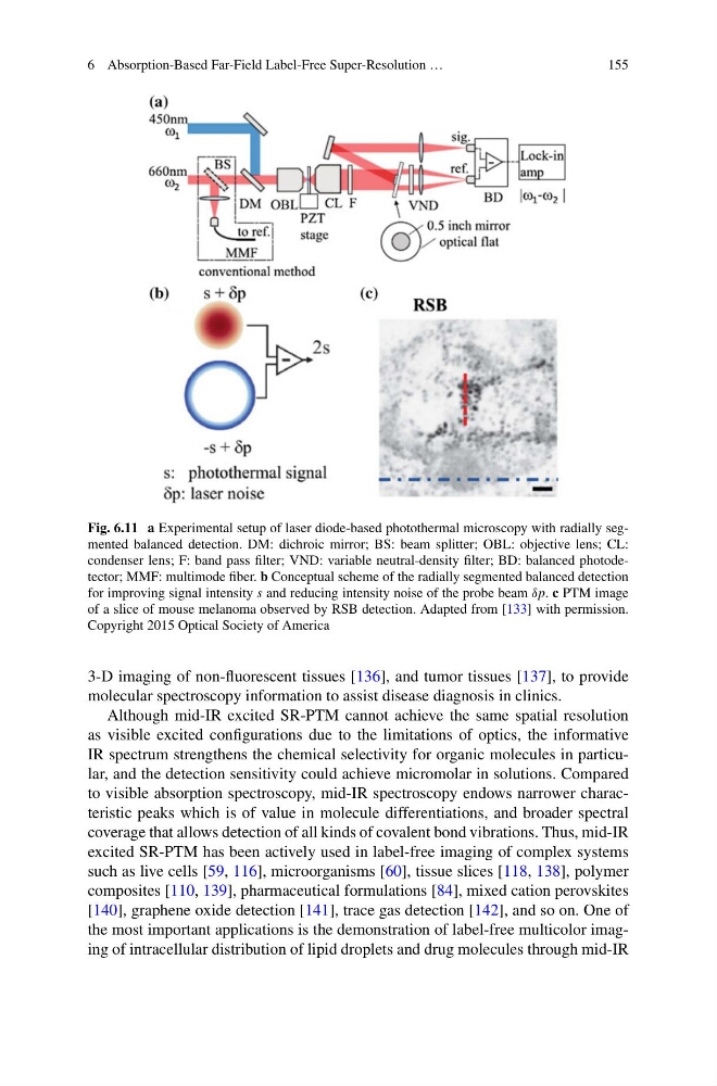
Fig. 6.11 a Experimental setup of laser diode-based photothermal microscopy with radially segmented balanced detection. DM: dichroic mirror; BS: beam splitter; OBL: objective lens; CL:
condenser lens; F: band pass filter; VND: variable neutral-density filter; BD: balanced photodetector; MMF: multimode fiber. b Conceptual scheme of the radially segmented balanced detection
for improving signal intensity s and reducing intensity noise of the probe beam δp. c PTM image
of a slice of mouse melanoma observed by RSB detection. Adapted from [133] with permission.
Copyright 2015 Optical Society of America
3-D imaging of non-fluorescent tissues [136], and tumor tissues [137], to provide
molecular spectroscopy information to assist disease diagnosis in clinics.
Although mid-IR excited SR-PTM cannot achieve the same spatial resolution
as visible excited configurations due to the limitations of optics, the informative
IR spectrum strengthens the chemical selectivity for organic molecules in particular, and the detection sensitivity could achieve micromolar in solutions. Compared
to visible absorption spectroscopy, mid-IR spectroscopy endows narrower characteristic peaks which is of value in molecule differentiations, and broader spectral
coverage that allows detection of all kinds of covalent bond vibrations. Thus, mid-IR
excited SR-PTM has been actively used in label-free imaging of complex systems
such as live cells [59, 116], microorganisms [60], tissue slices [118, 138], polymer
composites [110, 139], pharmaceutical formulations [84], mixed cation perovskites
[140], graphene oxide detection [141], trace gas detection [142], and so on. One of
the most important applications is the demonstration of label-free multicolor imaging of intracellular distribution of lipid droplets and drug molecules through mid-IR
155
Fig. 6.11 a Experimental setup of laser diode-based photothermal microscopy with radially segmented balanced detection. DM: dichroic mirror; BS: beam splitter; OBL: objective lens; CL:
condenser lens; F: band pass filter; VND: variable neutral-density filter; BD: balanced photodetector; MMF: multimode fiber. b Conceptual scheme of the radially segmented balanced detection
for improving signal intensity s and reducing intensity noise of the probe beam δp. c PTM image
of a slice of mouse melanoma observed by RSB detection. Adapted from [133] with permission.
Copyright 2015 Optical Society of America
3-D imaging of non-fluorescent tissues [136], and tumor tissues [137], to provide
molecular spectroscopy information to assist disease diagnosis in clinics.
Although mid-IR excited SR-PTM cannot achieve the same spatial resolution
as visible excited configurations due to the limitations of optics, the informative
IR spectrum strengthens the chemical selectivity for organic molecules in particular, and the detection sensitivity could achieve micromolar in solutions. Compared
to visible absorption spectroscopy, mid-IR spectroscopy endows narrower characteristic peaks which is of value in molecule differentiations, and broader spectral
coverage that allows detection of all kinds of covalent bond vibrations. Thus, mid-IR
excited SR-PTM has been actively used in label-free imaging of complex systems
such as live cells [59, 116], microorganisms [60], tissue slices [118, 138], polymer
composites [110, 139], pharmaceutical formulations [84], mixed cation perovskites
[140], graphene oxide detection [141], trace gas detection [142], and so on. One of
the most important applications is the demonstration of label-free multicolor imaging of intracellular distribution of lipid droplets and drug molecules through mid-IR