

146
C. Li and J.-X. Cheng
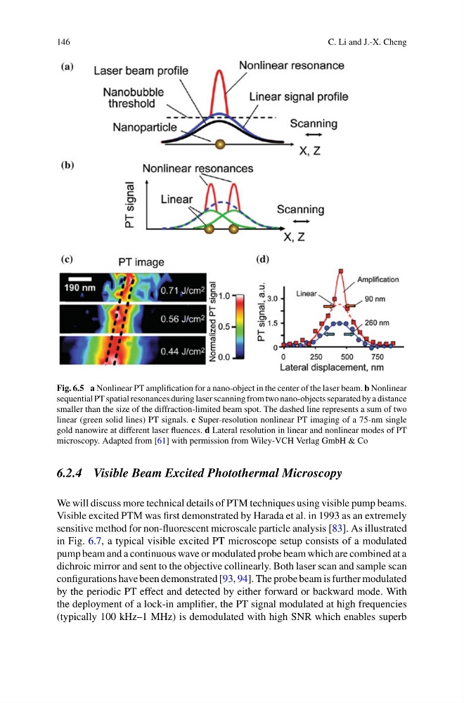
Fig. 6.5 a Nonlinear PT amplification for a nano-object in the center of the laser beam. b Nonlinear
sequential PT spatial resonances during laser scanning from two nano-objects separated by a distance
smaller than the size of the diffraction-limited beam spot. The dashed line represents a sum of two
linear (green solid lines) PT signals. c Super-resolution nonlinear PT imaging of a 75-nm single
gold nanowire at different laser fluences. d Lateral resolution in linear and nonlinear modes of PT
microscopy. Adapted from [61] with permission from Wiley-VCH Verlag GmbH & Co
6.2.4 Visible Beam Excited Photothermal Microscopy
We will discuss more technical details of PTM techniques using visible pump beams.
Visible excited PTM was first demonstrated by Harada et al. in 1993 as an extremely
sensitive method for non-fluorescent microscale particle analysis [83]. As illustrated
in Fig. 6.7, a typical visible excited PT microscope setup consists of a modulated
pump beam and a continuous wave or modulated probe beam which are combined at a
dichroic mirror and sent to the objective collinearly. Both laser scan and sample scan
configurations have been demonstrated [93, 94]. The probe beam is further modulated
by the periodic PT effect and detected by either forward or backward mode. With
the deployment of a lock-in amplifier, the PT signal modulated at high frequencies
(typically 100 kHz–1 MHz) is demodulated with high SNR which enables superb
C. Li and J.-X. Cheng
Fig. 6.5 a Nonlinear PT amplification for a nano-object in the center of the laser beam. b Nonlinear
sequential PT spatial resonances during laser scanning from two nano-objects separated by a distance
smaller than the size of the diffraction-limited beam spot. The dashed line represents a sum of two
linear (green solid lines) PT signals. c Super-resolution nonlinear PT imaging of a 75-nm single
gold nanowire at different laser fluences. d Lateral resolution in linear and nonlinear modes of PT
microscopy. Adapted from [61] with permission from Wiley-VCH Verlag GmbH & Co
6.2.4 Visible Beam Excited Photothermal Microscopy
We will discuss more technical details of PTM techniques using visible pump beams.
Visible excited PTM was first demonstrated by Harada et al. in 1993 as an extremely
sensitive method for non-fluorescent microscale particle analysis [83]. As illustrated
in Fig. 6.7, a typical visible excited PT microscope setup consists of a modulated
pump beam and a continuous wave or modulated probe beam which are combined at a
dichroic mirror and sent to the objective collinearly. Both laser scan and sample scan
configurations have been demonstrated [93, 94]. The probe beam is further modulated
by the periodic PT effect and detected by either forward or backward mode. With
the deployment of a lock-in amplifier, the PT signal modulated at high frequencies
(typically 100 kHz–1 MHz) is demodulated with high SNR which enables superb