

6 Absorption-Based Far-Field Label-Free Super-Resolution …
147
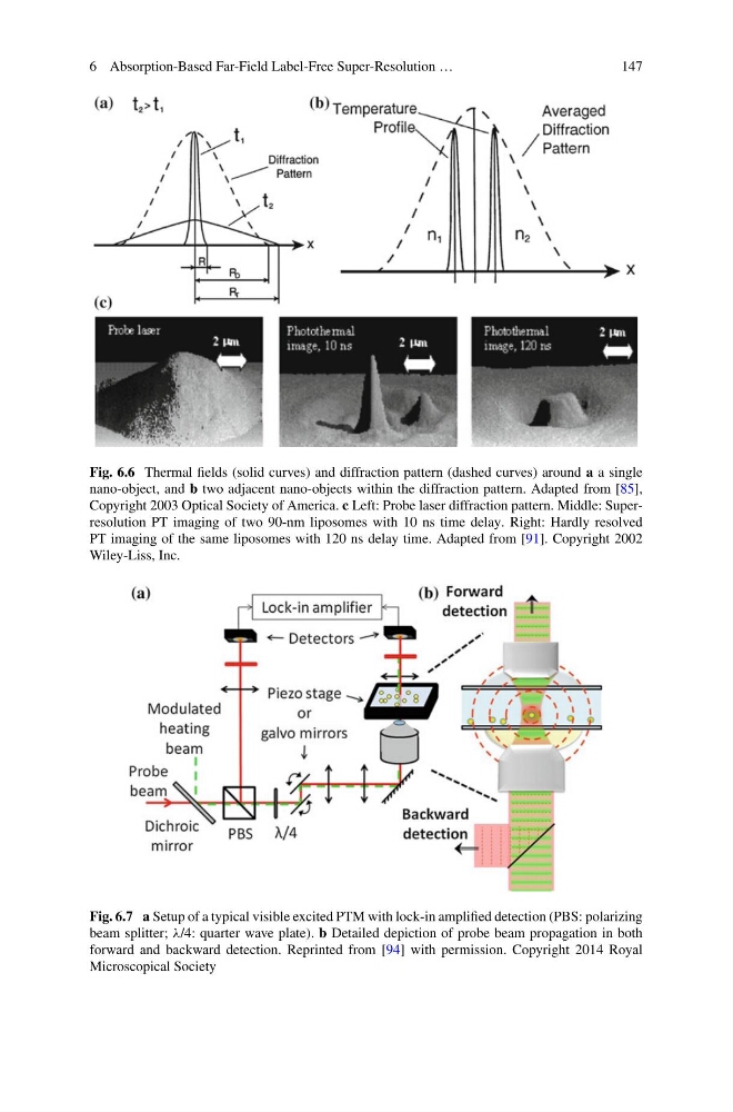
Fig. 6.6 Thermal fields (solid curves) and diffraction pattern (dashed curves) around a a single
nano-object, and b two adjacent nano-objects within the diffraction pattern. Adapted from [85],
Copyright 2003 Optical Society of America. c Left: Probe laser diffraction pattern. Middle: Superresolution PT imaging of two 90-nm liposomes with 10 ns time delay. Right: Hardly resolved
PT imaging of the same liposomes with 120 ns delay time. Adapted from [91]. Copyright 2002
Wiley-Liss, Inc.
Fig. 6.7 a Setup of a typical visible excited PTM with lock-in amplified detection (PBS: polarizing
beam splitter; λ/4: quarter wave plate). b Detailed depiction of probe beam propagation in both
forward and backward detection. Reprinted from [94] with permission. Copyright 2014 Royal
Microscopical Society
147
Fig. 6.6 Thermal fields (solid curves) and diffraction pattern (dashed curves) around a a single
nano-object, and b two adjacent nano-objects within the diffraction pattern. Adapted from [85],
Copyright 2003 Optical Society of America. c Left: Probe laser diffraction pattern. Middle: Superresolution PT imaging of two 90-nm liposomes with 10 ns time delay. Right: Hardly resolved
PT imaging of the same liposomes with 120 ns delay time. Adapted from [91]. Copyright 2002
Wiley-Liss, Inc.
Fig. 6.7 a Setup of a typical visible excited PTM with lock-in amplified detection (PBS: polarizing
beam splitter; λ/4: quarter wave plate). b Detailed depiction of probe beam propagation in both
forward and backward detection. Reprinted from [94] with permission. Copyright 2014 Royal
Microscopical Society