

6 Absorption-Based Far-Field Label-Free Super-Resolution …
145
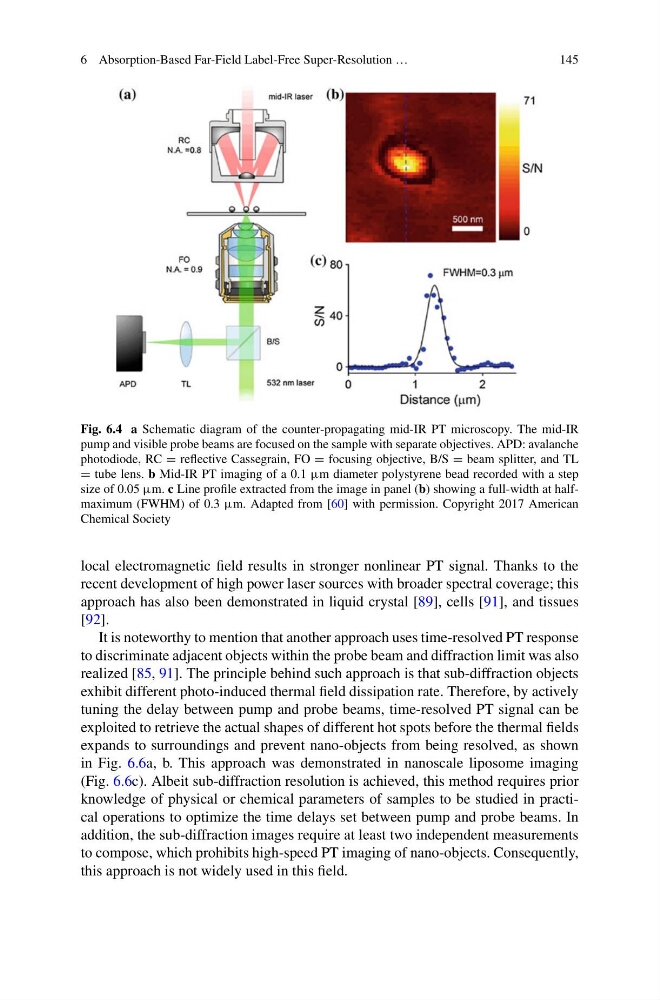
Fig. 6.4 a Schematic diagram of the counter-propagating mid-IR PT microscopy. The mid-IR
pump and visible probe beams are focused on the sample with separate objectives. APD: avalanche
photodiode, RC = reflective Cassegrain, FO = focusing objective, B/S = beam splitter, and TL
= tube lens. b Mid-IR PT imaging of a 0.1 µm diameter polystyrene bead recorded with a step
size of 0.05 µm. c Line profile extracted from the image in panel (b) showing a full-width at halfmaximum (FWHM) of 0.3 µm. Adapted from [60] with permission. Copyright 2017 American
Chemical Society
local electromagnetic field results in stronger nonlinear PT signal. Thanks to the
recent development of high power laser sources with broader spectral coverage; this
approach has also been demonstrated in liquid crystal [89], cells [91], and tissues
[92].
It is noteworthy to mention that another approach uses time-resolved PT response
to discriminate adjacent objects within the probe beam and diffraction limit was also
realized [85, 91]. The principle behind such approach is that sub-diffraction objects
exhibit different photo-induced thermal field dissipation rate. Therefore, by actively
tuning the delay between pump and probe beams, time-resolved PT signal can be
exploited to retrieve the actual shapes of different hot spots before the thermal fields
expands to surroundings and prevent nano-objects from being resolved, as shown
in Fig. 6.6a, b. This approach was demonstrated in nanoscale liposome imaging
(Fig. 6.6c). Albeit sub-diffraction resolution is achieved, this method requires prior
knowledge of physical or chemical parameters of samples to be studied in practical operations to optimize the time delays set between pump and probe beams. In
addition, the sub-diffraction images require at least two independent measurements
to compose, which prohibits high-speed PT imaging of nano-objects. Consequently,
this approach is not widely used in this field.
145
Fig. 6.4 a Schematic diagram of the counter-propagating mid-IR PT microscopy. The mid-IR
pump and visible probe beams are focused on the sample with separate objectives. APD: avalanche
photodiode, RC = reflective Cassegrain, FO = focusing objective, B/S = beam splitter, and TL
= tube lens. b Mid-IR PT imaging of a 0.1 µm diameter polystyrene bead recorded with a step
size of 0.05 µm. c Line profile extracted from the image in panel (b) showing a full-width at halfmaximum (FWHM) of 0.3 µm. Adapted from [60] with permission. Copyright 2017 American
Chemical Society
local electromagnetic field results in stronger nonlinear PT signal. Thanks to the
recent development of high power laser sources with broader spectral coverage; this
approach has also been demonstrated in liquid crystal [89], cells [91], and tissues
[92].
It is noteworthy to mention that another approach uses time-resolved PT response
to discriminate adjacent objects within the probe beam and diffraction limit was also
realized [85, 91]. The principle behind such approach is that sub-diffraction objects
exhibit different photo-induced thermal field dissipation rate. Therefore, by actively
tuning the delay between pump and probe beams, time-resolved PT signal can be
exploited to retrieve the actual shapes of different hot spots before the thermal fields
expands to surroundings and prevent nano-objects from being resolved, as shown
in Fig. 6.6a, b. This approach was demonstrated in nanoscale liposome imaging
(Fig. 6.6c). Albeit sub-diffraction resolution is achieved, this method requires prior
knowledge of physical or chemical parameters of samples to be studied in practical operations to optimize the time delays set between pump and probe beams. In
addition, the sub-diffraction images require at least two independent measurements
to compose, which prohibits high-speed PT imaging of nano-objects. Consequently,
this approach is not widely used in this field.