

98
B. Simon and O. Haeberlé
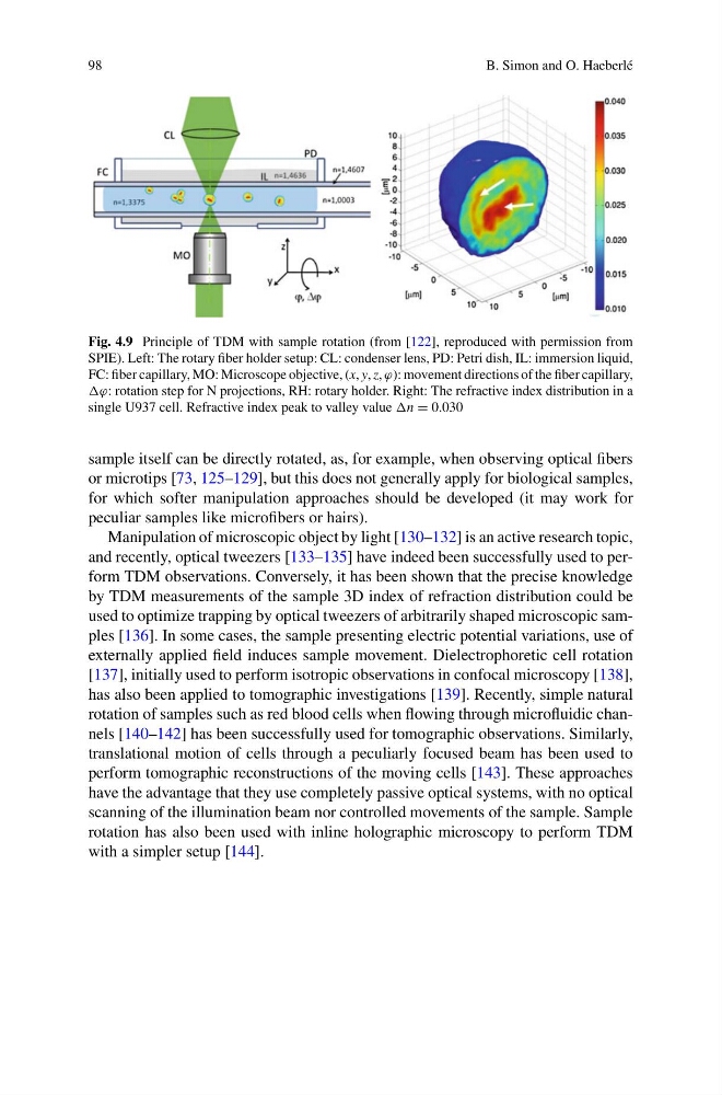
Fig. 4.9 Principle of TDM with sample rotation (from [122], reproduced with permission from
SPIE). Left: The rotary fiber holder setup: CL: condenser lens, PD: Petri dish, IL: immersion liquid,
FC: fiber capillary, MO: Microscope objective, (x, y, z, ϕ): movement directions of the fiber capillary,
ϕ: rotation step for N projections, RH: rotary holder. Right: The refractive index distribution in a
single U937 cell. Refractive index peak to valley value n = 0.030
sample itself can be directly rotated, as, for example, when observing optical fibers
or microtips [73, 125–129], but this does not generally apply for biological samples,
for which softer manipulation approaches should be developed (it may work for
peculiar samples like microfibers or hairs).
Manipulation of microscopic object by light [130–132] is an active research topic,
and recently, optical tweezers [133–135] have indeed been successfully used to perform TDM observations. Conversely, it has been shown that the precise knowledge
by TDM measurements of the sample 3D index of refraction distribution could be
used to optimize trapping by optical tweezers of arbitrarily shaped microscopic samples [136]. In some cases, the sample presenting electric potential variations, use of
externally applied field induces sample movement. Dielectrophoretic cell rotation
[137], initially used to perform isotropic observations in confocal microscopy [138],
has also been applied to tomographic investigations [139]. Recently, simple natural
rotation of samples such as red blood cells when flowing through microfluidic channels [140–142] has been successfully used for tomographic observations. Similarly,
translational motion of cells through a peculiarly focused beam has been used to
perform tomographic reconstructions of the moving cells [143]. These approaches
have the advantage that they use completely passive optical systems, with no optical
scanning of the illumination beam nor controlled movements of the sample. Sample
rotation has also been used with inline holographic microscopy to perform TDM
with a simpler setup [144].
B. Simon and O. Haeberlé
Fig. 4.9 Principle of TDM with sample rotation (from [122], reproduced with permission from
SPIE). Left: The rotary fiber holder setup: CL: condenser lens, PD: Petri dish, IL: immersion liquid,
FC: fiber capillary, MO: Microscope objective, (x, y, z, ϕ): movement directions of the fiber capillary,
ϕ: rotation step for N projections, RH: rotary holder. Right: The refractive index distribution in a
single U937 cell. Refractive index peak to valley value n = 0.030
sample itself can be directly rotated, as, for example, when observing optical fibers
or microtips [73, 125–129], but this does not generally apply for biological samples,
for which softer manipulation approaches should be developed (it may work for
peculiar samples like microfibers or hairs).
Manipulation of microscopic object by light [130–132] is an active research topic,
and recently, optical tweezers [133–135] have indeed been successfully used to perform TDM observations. Conversely, it has been shown that the precise knowledge
by TDM measurements of the sample 3D index of refraction distribution could be
used to optimize trapping by optical tweezers of arbitrarily shaped microscopic samples [136]. In some cases, the sample presenting electric potential variations, use of
externally applied field induces sample movement. Dielectrophoretic cell rotation
[137], initially used to perform isotropic observations in confocal microscopy [138],
has also been applied to tomographic investigations [139]. Recently, simple natural
rotation of samples such as red blood cells when flowing through microfluidic channels [140–142] has been successfully used for tomographic observations. Similarly,
translational motion of cells through a peculiarly focused beam has been used to
perform tomographic reconstructions of the moving cells [143]. These approaches
have the advantage that they use completely passive optical systems, with no optical
scanning of the illumination beam nor controlled movements of the sample. Sample
rotation has also been used with inline holographic microscopy to perform TDM
with a simpler setup [144].