

94
B. Simon and O. Haeberlé
depicts human white blood cells images with TDM, highlighting its ability to segment
membrane from internal structures, without any specific labeling.
Commercial implementations of TDM with illumination rotation are now available. See the websites of Nanolive [106] and Tomocube [107] companies for various
examples of applications on biological samples. The nanolive system has the advantage to offer a large accessible space over the sample, while the Tomocube allows
for faster acquisitions at slightly higher resolutions.
4.4 Multiwavelength TDM
Tomographic Diffractive Microscopy with illumination rotation is now developed
in several laboratories, and even commercially available, but requires specific illumination systems to perform numerical aperture synthesis of the condenser. This
can be performed onto a standard microscope body [67, 83–85], but explains why
specific setups are often preferred for laboratory prototyping, as well as for commercial implementations [106, 107]. This has then the drawback that TDM cannot
be performed using a standard widefield or confocal fluorescence microscope, often
already available in biological laboratories.
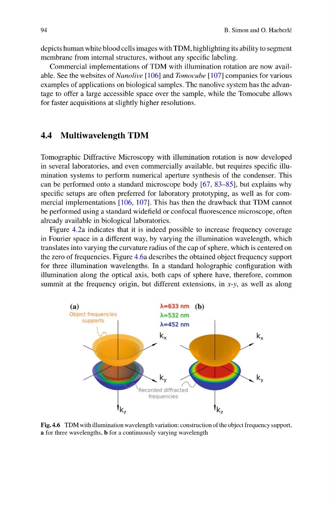
Figure 4.2a indicates that it is indeed possible to increase frequency coverage
in Fourier space in a different way, by varying the illumination wavelength, which
translates into varying the curvature radius of the cap of sphere, which is centered on
the zero of frequencies. Figure 4.6a describes the obtained object frequency support
for three illumination wavelengths. In a standard holographic configuration with
illumination along the optical axis, both caps of sphere have, therefore, common
summit at the frequency origin, but different extensions, in x-y, as well as along
Fig. 4.6 TDM with illumination wavelength variation: construction of the object frequency support,
a for three wavelengths, b for a continuously varying wavelength
B. Simon and O. Haeberlé
depicts human white blood cells images with TDM, highlighting its ability to segment
membrane from internal structures, without any specific labeling.
Commercial implementations of TDM with illumination rotation are now available. See the websites of Nanolive [106] and Tomocube [107] companies for various
examples of applications on biological samples. The nanolive system has the advantage to offer a large accessible space over the sample, while the Tomocube allows
for faster acquisitions at slightly higher resolutions.
4.4 Multiwavelength TDM
Tomographic Diffractive Microscopy with illumination rotation is now developed
in several laboratories, and even commercially available, but requires specific illumination systems to perform numerical aperture synthesis of the condenser. This
can be performed onto a standard microscope body [67, 83–85], but explains why
specific setups are often preferred for laboratory prototyping, as well as for commercial implementations [106, 107]. This has then the drawback that TDM cannot
be performed using a standard widefield or confocal fluorescence microscope, often
already available in biological laboratories.
Figure 4.2a indicates that it is indeed possible to increase frequency coverage
in Fourier space in a different way, by varying the illumination wavelength, which
translates into varying the curvature radius of the cap of sphere, which is centered on
the zero of frequencies. Figure 4.6a describes the obtained object frequency support
for three illumination wavelengths. In a standard holographic configuration with
illumination along the optical axis, both caps of sphere have, therefore, common
summit at the frequency origin, but different extensions, in x-y, as well as along
Fig. 4.6 TDM with illumination wavelength variation: construction of the object frequency support,
a for three wavelengths, b for a continuously varying wavelength