

92
B. Simon and O. Haeberlé
So, in order to improve image quality, one has to expand the k o coverage. In TDM
with illumination rotation, the illumination can be tilted (Fig. 4.2b). In that case, one
can finally record a complementary subset of information (Fig. 4.2c).
For a large number of illuminations, a synthetic aperture approach delivers an
enlarged frequency support. In that configuration, one recognizes the characteristic “doughnut” shape of transmission optical microscopy [82]. This extended frequency support explains the better resolution and optical sectioning capability of
TDM, compared to DHM. On can theoretically double the lateral resolution [78,
81], experimentally reaching resolution in the 100 nm range [83].
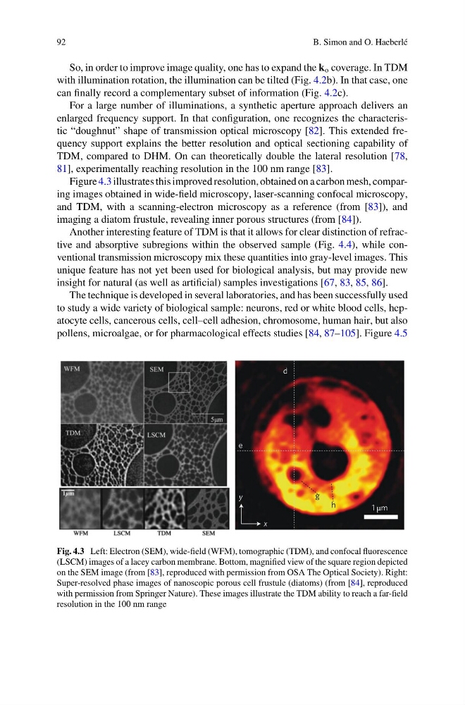
Figure 4.3 illustrates this improved resolution, obtained on a carbon mesh, comparing images obtained in wide-field microscopy, laser-scanning confocal microscopy,
and TDM, with a scanning-electron microscopy as a reference (from [83]), and
imaging a diatom frustule, revealing inner porous structures (from [84]).
Another interesting feature of TDM is that it allows for clear distinction of refractive and absorptive subregions within the observed sample (Fig. 4.4), while conventional transmission microscopy mix these quantities into gray-level images. This
unique feature has not yet been used for biological analysis, but may provide new
insight for natural (as well as artificial) samples investigations [67, 83, 85, 86].
The technique is developed in several laboratories, and has been successfully used
to study a wide variety of biological sample: neurons, red or white blood cells, hepatocyte cells, cancerous cells, cell–cell adhesion, chromosome, human hair, but also
pollens, microalgae, or for pharmacological effects studies [84, 87–105]. Figure 4.5
Fig. 4.3 Left: Electron (SEM), wide-field (WFM), tomographic (TDM), and confocal fluorescence
(LSCM) images of a lacey carbon membrane. Bottom, magnified view of the square region depicted
on the SEM image (from [83], reproduced with permission from OSA The Optical Society). Right:
Super-resolved phase images of nanoscopic porous cell frustule (diatoms) (from [84], reproduced
with permission from Springer Nature). These images illustrate the TDM ability to reach a far-field
resolution in the 100 nm range
B. Simon and O. Haeberlé
So, in order to improve image quality, one has to expand the k o coverage. In TDM
with illumination rotation, the illumination can be tilted (Fig. 4.2b). In that case, one
can finally record a complementary subset of information (Fig. 4.2c).
For a large number of illuminations, a synthetic aperture approach delivers an
enlarged frequency support. In that configuration, one recognizes the characteristic “doughnut” shape of transmission optical microscopy [82]. This extended frequency support explains the better resolution and optical sectioning capability of
TDM, compared to DHM. On can theoretically double the lateral resolution [78,
81], experimentally reaching resolution in the 100 nm range [83].
Figure 4.3 illustrates this improved resolution, obtained on a carbon mesh, comparing images obtained in wide-field microscopy, laser-scanning confocal microscopy,
and TDM, with a scanning-electron microscopy as a reference (from [83]), and
imaging a diatom frustule, revealing inner porous structures (from [84]).
Another interesting feature of TDM is that it allows for clear distinction of refractive and absorptive subregions within the observed sample (Fig. 4.4), while conventional transmission microscopy mix these quantities into gray-level images. This
unique feature has not yet been used for biological analysis, but may provide new
insight for natural (as well as artificial) samples investigations [67, 83, 85, 86].
The technique is developed in several laboratories, and has been successfully used
to study a wide variety of biological sample: neurons, red or white blood cells, hepatocyte cells, cancerous cells, cell–cell adhesion, chromosome, human hair, but also
pollens, microalgae, or for pharmacological effects studies [84, 87–105]. Figure 4.5
Fig. 4.3 Left: Electron (SEM), wide-field (WFM), tomographic (TDM), and confocal fluorescence
(LSCM) images of a lacey carbon membrane. Bottom, magnified view of the square region depicted
on the SEM image (from [83], reproduced with permission from OSA The Optical Society). Right:
Super-resolved phase images of nanoscopic porous cell frustule (diatoms) (from [84], reproduced
with permission from Springer Nature). These images illustrate the TDM ability to reach a far-field
resolution in the 100 nm range