

4 Tomographic Diffractive Microscopy …
91
The scattered far-field amplitude is written as
e(k) = k
2
0
exp
−ik · r
X
r
E
r
dr
(4.5)
Considering a plane wave with wavevector k inc as incident field, the incident field
is written as E inc (r) = A inc exp(ik inc · r), and limiting the development to a weakly
diffracting object, for which the first Born approximation holds, one can finally write
e(k, k inc ) = C ˜
X (k − k inc )
(4.6)
In (4.6), C = 8π
3 A inc k
2
0 . Equation (4.6) provides a direct correspondence between
the diffracted far-field amplitude and the Fourier coefficient of the relative permittivity of the object.
Energy conservation in the diffraction process implies that diffracted k diff vectors
have same amplitude as k inc , and therefore depict the so-called Ewald sphere. Because
of the limited numerical aperture of the detection objective, only a cap of the Ewald
sphere can be captured.
Then from the elastic scattering condition, this set of diffracted k diff vectors transforms into object k o vectors, via a simple translation:
k o = k diff − k inc
(4.7)
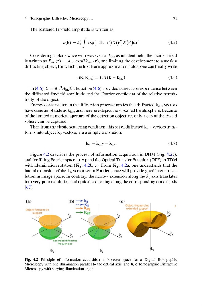
Figure 4.2 describes the process of information acquisition in DHM (Fig. 4.2a),
and for filling Fourier space to expand the Optical Transfer Function (OTF) in TDM
with illumination rotation (Fig. 4.2b, c). From Fig. 4.2a, one understands that the
lateral extension of the k o vector set in Fourier space will provide good lateral resolution in image space. In contrary, the narrow extension along the k z axis translates
into very poor resolution and optical sectioning along the corresponding optical axis
[67].
Fig. 4.2 Principle of information acquisition in k-vector space for a Digital Holographic
Microscopy with one illumination parallel to the optical axis, and b, c Tomographic Diffractive
Microscopy with varying illumination angle
91
The scattered far-field amplitude is written as
e(k) = k
2
0
exp
−ik · r
X
r
E
r
dr
(4.5)
Considering a plane wave with wavevector k inc as incident field, the incident field
is written as E inc (r) = A inc exp(ik inc · r), and limiting the development to a weakly
diffracting object, for which the first Born approximation holds, one can finally write
e(k, k inc ) = C ˜
X (k − k inc )
(4.6)
In (4.6), C = 8π
3 A inc k
2
0 . Equation (4.6) provides a direct correspondence between
the diffracted far-field amplitude and the Fourier coefficient of the relative permittivity of the object.
Energy conservation in the diffraction process implies that diffracted k diff vectors
have same amplitude as k inc , and therefore depict the so-called Ewald sphere. Because
of the limited numerical aperture of the detection objective, only a cap of the Ewald
sphere can be captured.
Then from the elastic scattering condition, this set of diffracted k diff vectors transforms into object k o vectors, via a simple translation:
k o = k diff − k inc
(4.7)
Figure 4.2 describes the process of information acquisition in DHM (Fig. 4.2a),
and for filling Fourier space to expand the Optical Transfer Function (OTF) in TDM
with illumination rotation (Fig. 4.2b, c). From Fig. 4.2a, one understands that the
lateral extension of the k o vector set in Fourier space will provide good lateral resolution in image space. In contrary, the narrow extension along the k z axis translates
into very poor resolution and optical sectioning along the corresponding optical axis
[67].
Fig. 4.2 Principle of information acquisition in k-vector space for a Digital Holographic
Microscopy with one illumination parallel to the optical axis, and b, c Tomographic Diffractive
Microscopy with varying illumination angle