

13 Computational Biomechanics of the Brain in the Operating Theatre
341
Pre-operative Steps
Segmentation of the pre-operative MRI remains one of the main limitations for a
widespread use of our method in clinic. Indeed, while the whole brain is extracted
via the automatic BET algorithm [9], the segmentation of the tumour and the
other anatomical structures remains manual. Nevertheless, the segmentation of preoperative images tends to become a standard step of nowadays clinical routines.
Moreover, several solutions exist in literature [23–25] for automatic extraction of
the brain and its various anatomical structures from MRIs.
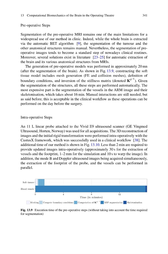
The generation of pre-operative models was performed in approximately 20 mn
(after the segmentation of the brain). As shown in Fig. 13.9, constructing the soft
tissue model includes mesh generation (FE and collision meshes), definition of
boundary conditions, and inversion of the stiffness matrix (denoted K 0 −1 ). Given
the segmentation of the structures, all these steps are performed automatically. The
most expensive part is the segmentation of the vessels in the ARM image and their
skeletonisation, which takes about 16 min. Manual interactions are still needed, but
as said before, this is acceptable in the clinical workflow as these operations can be
performed on the day before the surgery.
Intra-operative Steps
An 11 L linear probe attached to the Vivid E9 ultrasound scanner (GE Vingmed
Ultrasound, Horten, Norway) was used for all acquisitions. The 3D reconstruction of
images and the initial rigid transformation were performed intra-operatively with the
CustusX framework, which was successfully used in a clinical workflow [38]. The
additional time of our method is shown in Fig. 13.10. Less than 2 min are required to
provide updated images intra-operatively (approximately 30 s for the extraction of
vessels and the footprint, 1–2 mm for the simulation and 10 s to warp the image). In
addition, the mode B and Doppler ultrasound images being acquired simultaneously,
the extraction of the footprint of the probe, and the vessels can be performed in
parallel.
Blood vessels
Soft tissues
0
4
8
1 2
Time (in minutes)
Meshing
Compute boundary conditions
Computation of K
0 −1
MIP segmentation
Skeletonisation
Fig. 13.9 Execution time of the pre-operative steps (without taking into account the time required
for segmentation)
341
Pre-operative Steps
Segmentation of the pre-operative MRI remains one of the main limitations for a
widespread use of our method in clinic. Indeed, while the whole brain is extracted
via the automatic BET algorithm [9], the segmentation of the tumour and the
other anatomical structures remains manual. Nevertheless, the segmentation of preoperative images tends to become a standard step of nowadays clinical routines.
Moreover, several solutions exist in literature [23–25] for automatic extraction of
the brain and its various anatomical structures from MRIs.
The generation of pre-operative models was performed in approximately 20 mn
(after the segmentation of the brain). As shown in Fig. 13.9, constructing the soft
tissue model includes mesh generation (FE and collision meshes), definition of
boundary conditions, and inversion of the stiffness matrix (denoted K 0 −1 ). Given
the segmentation of the structures, all these steps are performed automatically. The
most expensive part is the segmentation of the vessels in the ARM image and their
skeletonisation, which takes about 16 min. Manual interactions are still needed, but
as said before, this is acceptable in the clinical workflow as these operations can be
performed on the day before the surgery.
Intra-operative Steps
An 11 L linear probe attached to the Vivid E9 ultrasound scanner (GE Vingmed
Ultrasound, Horten, Norway) was used for all acquisitions. The 3D reconstruction of
images and the initial rigid transformation were performed intra-operatively with the
CustusX framework, which was successfully used in a clinical workflow [38]. The
additional time of our method is shown in Fig. 13.10. Less than 2 min are required to
provide updated images intra-operatively (approximately 30 s for the extraction of
vessels and the footprint, 1–2 mm for the simulation and 10 s to warp the image). In
addition, the mode B and Doppler ultrasound images being acquired simultaneously,
the extraction of the footprint of the probe, and the vessels can be performed in
parallel.
Blood vessels
Soft tissues
0
4
8
1 2
Time (in minutes)
Meshing
Compute boundary conditions
Computation of K
0 −1
MIP segmentation
Skeletonisation
Fig. 13.9 Execution time of the pre-operative steps (without taking into account the time required
for segmentation)