

340
H. Courtecuisse et al.
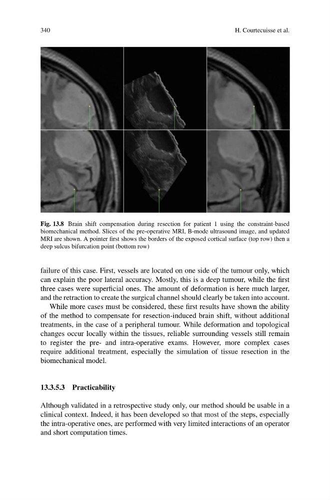
Fig. 13.8 Brain shift compensation during resection for patient 1 using the constraint-based
biomechanical method. Slices of the pre-operative MRI, B-mode ultrasound image, and updated
MRI are shown. A pointer first shows the borders of the exposed cortical surface (top row) then a
deep sulcus bifurcation point (bottom row)
failure of this case. First, vessels are located on one side of the tumour only, which
can explain the poor lateral accuracy. Mostly, this is a deep tumour, while the first
three cases were superficial ones. The amount of deformation is here much larger,
and the retraction to create the surgical channel should clearly be taken into account.
While more cases must be considered, these first results have shown the ability
of the method to compensate for resection-induced brain shift, without additional
treatments, in the case of a peripheral tumour. While deformation and topological
changes occur locally within the tissues, reliable surrounding vessels still remain
to register the pre- and intra-operative exams. However, more complex cases
require additional treatment, especially the simulation of tissue resection in the
biomechanical model.
13.3.5.3 Practicability
Although validated in a retrospective study only, our method should be usable in a
clinical context. Indeed, it has been developed so that most of the steps, especially
the intra-operative ones, are performed with very limited interactions of an operator
and short computation times.
H. Courtecuisse et al.
Fig. 13.8 Brain shift compensation during resection for patient 1 using the constraint-based
biomechanical method. Slices of the pre-operative MRI, B-mode ultrasound image, and updated
MRI are shown. A pointer first shows the borders of the exposed cortical surface (top row) then a
deep sulcus bifurcation point (bottom row)
failure of this case. First, vessels are located on one side of the tumour only, which
can explain the poor lateral accuracy. Mostly, this is a deep tumour, while the first
three cases were superficial ones. The amount of deformation is here much larger,
and the retraction to create the surgical channel should clearly be taken into account.
While more cases must be considered, these first results have shown the ability
of the method to compensate for resection-induced brain shift, without additional
treatments, in the case of a peripheral tumour. While deformation and topological
changes occur locally within the tissues, reliable surrounding vessels still remain
to register the pre- and intra-operative exams. However, more complex cases
require additional treatment, especially the simulation of tissue resection in the
biomechanical model.
13.3.5.3 Practicability
Although validated in a retrospective study only, our method should be usable in a
clinical context. Indeed, it has been developed so that most of the steps, especially
the intra-operative ones, are performed with very limited interactions of an operator
and short computation times.