

13 Computational Biomechanics of the Brain in the Operating Theatre
339
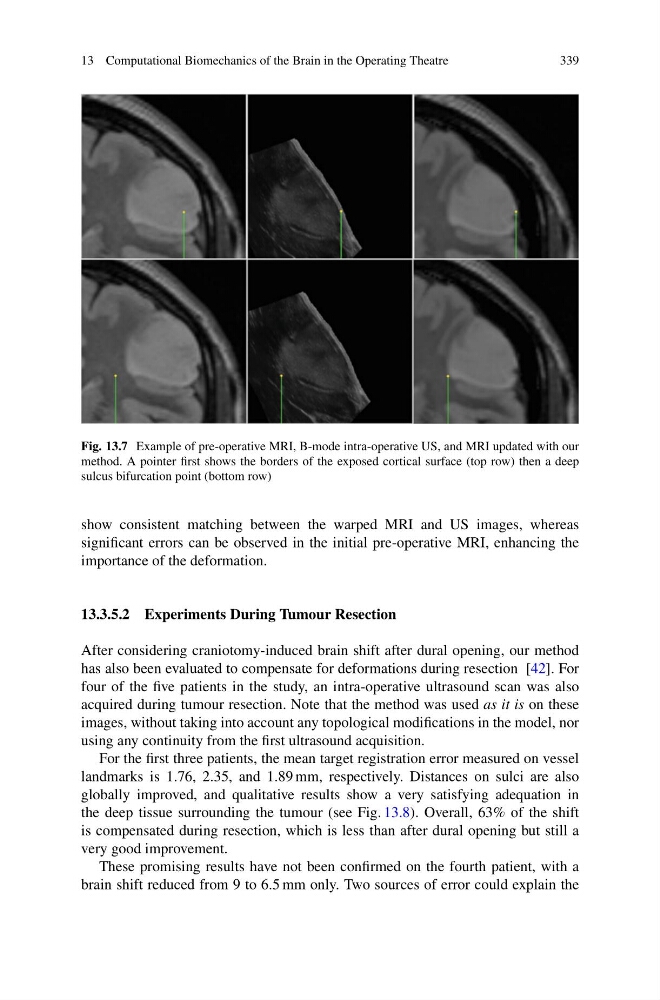
Fig. 13.7 Example of pre-operative MRI, B-mode intra-operative US, and MRI updated with our
method. A pointer first shows the borders of the exposed cortical surface (top row) then a deep
sulcus bifurcation point (bottom row)
show consistent matching between the warped MRI and US images, whereas
significant errors can be observed in the initial pre-operative MRI, enhancing the
importance of the deformation.
13.3.5.2 Experiments During Tumour Resection
After considering craniotomy-induced brain shift after dural opening, our method
has also been evaluated to compensate for deformations during resection [42]. For
four of the five patients in the study, an intra-operative ultrasound scan was also
acquired during tumour resection. Note that the method was used as it is on these
images, without taking into account any topological modifications in the model, nor
using any continuity from the first ultrasound acquisition.
For the first three patients, the mean target registration error measured on vessel
landmarks is 1.76, 2.35, and 1.89 mm, respectively. Distances on sulci are also
globally improved, and qualitative results show a very satisfying adequation in
the deep tissue surrounding the tumour (see Fig. 13.8). Overall, 63% of the shift
is compensated during resection, which is less than after dural opening but still a
very good improvement.
These promising results have not been confirmed on the fourth patient, with a
brain shift reduced from 9 to 6.5 mm only. Two sources of error could explain the
339
Fig. 13.7 Example of pre-operative MRI, B-mode intra-operative US, and MRI updated with our
method. A pointer first shows the borders of the exposed cortical surface (top row) then a deep
sulcus bifurcation point (bottom row)
show consistent matching between the warped MRI and US images, whereas
significant errors can be observed in the initial pre-operative MRI, enhancing the
importance of the deformation.
13.3.5.2 Experiments During Tumour Resection
After considering craniotomy-induced brain shift after dural opening, our method
has also been evaluated to compensate for deformations during resection [42]. For
four of the five patients in the study, an intra-operative ultrasound scan was also
acquired during tumour resection. Note that the method was used as it is on these
images, without taking into account any topological modifications in the model, nor
using any continuity from the first ultrasound acquisition.
For the first three patients, the mean target registration error measured on vessel
landmarks is 1.76, 2.35, and 1.89 mm, respectively. Distances on sulci are also
globally improved, and qualitative results show a very satisfying adequation in
the deep tissue surrounding the tumour (see Fig. 13.8). Overall, 63% of the shift
is compensated during resection, which is less than after dural opening but still a
very good improvement.
These promising results have not been confirmed on the fourth patient, with a
brain shift reduced from 9 to 6.5 mm only. Two sources of error could explain the