

342
H. Courtecuisse et al.
Probe footprint
Blood vessels
0.0
0.5
1.0
1.5
Time (in minutes)
Extraction of blood vessels
Extraction of probe footprint
Simulation
MRI update
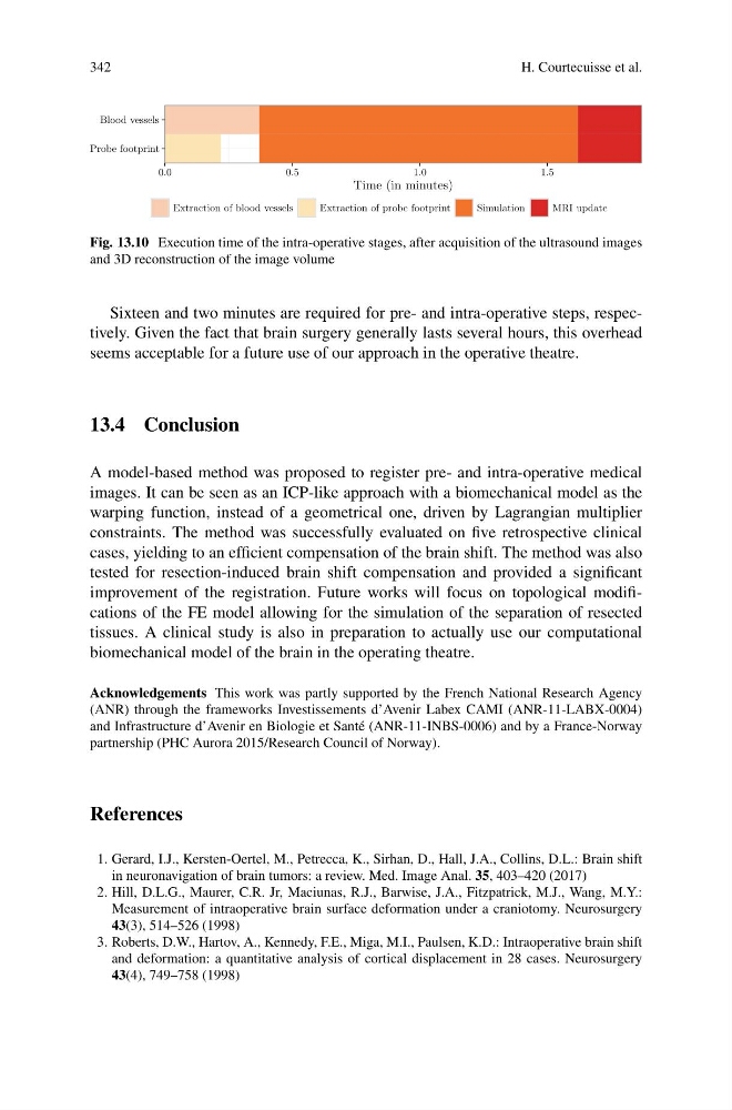
Fig. 13.10 Execution time of the intra-operative stages, after acquisition of the ultrasound images
and 3D reconstruction of the image volume
Sixteen and two minutes are required for pre- and intra-operative steps, respectively. Given the fact that brain surgery generally lasts several hours, this overhead
seems acceptable for a future use of our approach in the operative theatre.
13.4 Conclusion
A model-based method was proposed to register pre- and intra-operative medical
images. It can be seen as an ICP-like approach with a biomechanical model as the
warping function, instead of a geometrical one, driven by Lagrangian multiplier
constraints. The method was successfully evaluated on five retrospective clinical
cases, yielding to an efficient compensation of the brain shift. The method was also
tested for resection-induced brain shift compensation and provided a significant
improvement of the registration. Future works will focus on topological modifications of the FE model allowing for the simulation of the separation of resected
tissues. A clinical study is also in preparation to actually use our computational
biomechanical model of the brain in the operating theatre.
Acknowledgements This work was partly supported by the French National Research Agency
(ANR) through the frameworks Investissements d’Avenir Labex CAMI (ANR-11-LABX-0004)
and Infrastructure d’Avenir en Biologie et Santé (ANR-11-INBS-0006) and by a France-Norway
partnership (PHC Aurora 2015/Research Council of Norway).
References
1. Gerard, I.J., Kersten-Oertel, M., Petrecca, K., Sirhan, D., Hall, J.A., Collins, D.L.: Brain shift
in neuronavigation of brain tumors: a review. Med. Image Anal. 35, 403–420 (2017)
2. Hill, D.L.G., Maurer, C.R. Jr, Maciunas, R.J., Barwise, J.A., Fitzpatrick, M.J., Wang, M.Y.:
Measurement of intraoperative brain surface deformation under a craniotomy. Neurosurgery
43(3), 514–526 (1998)
3. Roberts, D.W., Hartov, A., Kennedy, F.E., Miga, M.I., Paulsen, K.D.: Intraoperative brain shift
and deformation: a quantitative analysis of cortical displacement in 28 cases. Neurosurgery
43(4), 749–758 (1998)
H. Courtecuisse et al.
Probe footprint
Blood vessels
0.0
0.5
1.0
1.5
Time (in minutes)
Extraction of blood vessels
Extraction of probe footprint
Simulation
MRI update
Fig. 13.10 Execution time of the intra-operative stages, after acquisition of the ultrasound images
and 3D reconstruction of the image volume
Sixteen and two minutes are required for pre- and intra-operative steps, respectively. Given the fact that brain surgery generally lasts several hours, this overhead
seems acceptable for a future use of our approach in the operative theatre.
13.4 Conclusion
A model-based method was proposed to register pre- and intra-operative medical
images. It can be seen as an ICP-like approach with a biomechanical model as the
warping function, instead of a geometrical one, driven by Lagrangian multiplier
constraints. The method was successfully evaluated on five retrospective clinical
cases, yielding to an efficient compensation of the brain shift. The method was also
tested for resection-induced brain shift compensation and provided a significant
improvement of the registration. Future works will focus on topological modifications of the FE model allowing for the simulation of the separation of resected
tissues. A clinical study is also in preparation to actually use our computational
biomechanical model of the brain in the operating theatre.
Acknowledgements This work was partly supported by the French National Research Agency
(ANR) through the frameworks Investissements d’Avenir Labex CAMI (ANR-11-LABX-0004)
and Infrastructure d’Avenir en Biologie et Santé (ANR-11-INBS-0006) and by a France-Norway
partnership (PHC Aurora 2015/Research Council of Norway).
References
1. Gerard, I.J., Kersten-Oertel, M., Petrecca, K., Sirhan, D., Hall, J.A., Collins, D.L.: Brain shift
in neuronavigation of brain tumors: a review. Med. Image Anal. 35, 403–420 (2017)
2. Hill, D.L.G., Maurer, C.R. Jr, Maciunas, R.J., Barwise, J.A., Fitzpatrick, M.J., Wang, M.Y.:
Measurement of intraoperative brain surface deformation under a craniotomy. Neurosurgery
43(3), 514–526 (1998)
3. Roberts, D.W., Hartov, A., Kennedy, F.E., Miga, M.I., Paulsen, K.D.: Intraoperative brain shift
and deformation: a quantitative analysis of cortical displacement in 28 cases. Neurosurgery
43(4), 749–758 (1998)