

206
L. Geregele et al.
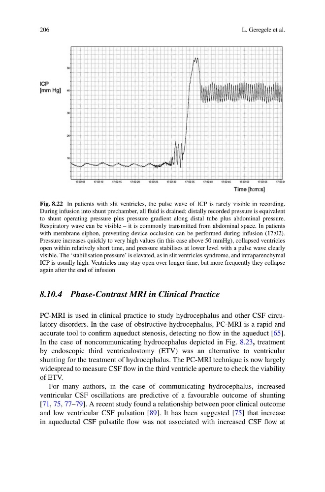
Fig. 8.22 In patients with slit ventricles, the pulse wave of ICP is rarely visible in recording.
During infusion into shunt prechamber, all fluid is drained; distally recorded pressure is equivalent
to shunt operating pressure plus pressure gradient along distal tube plus abdominal pressure.
Respiratory wave can be visible – it is commonly transmitted from abdominal space. In patients
with membrane siphon, preventing device occlusion can be performed during infusion (17:02).
Pressure increases quickly to very high values (in this case above 50 mmHg), collapsed ventricles
open within relatively short time, and pressure stabilises at lower level with a pulse wave clearly
visible. The ‘stabilisation pressure’ is elevated, as in slit ventricles syndrome, and intraparenchymal
ICP is usually high. Ventricles may stay open over longer time, but more frequently they collapse
again after the end of infusion
8.10.4 Phase-Contrast MRI in Clinical Practice
PC-MRI is used in clinical practice to study hydrocephalus and other CSF circulatory disorders. In the case of obstructive hydrocephalus, PC-MRI is a rapid and
accurate tool to confirm aqueduct stenosis, detecting no flow in the aqueduct [65].
In the case of noncommunicating hydrocephalus depicted in Fig. 8.23, treatment
by endoscopic third ventriculostomy (ETV) was an alternative to ventricular
shunting for the treatment of hydrocephalus. The PC-MRI technique is now largely
widespread to measure CSF flow in the third ventricle aperture to check the viability
of ETV.
For many authors, in the case of communicating hydrocephalus, increased
ventricular CSF oscillations are predictive of a favourable outcome of shunting
[71, 75, 77–79]. A recent study found a relationship between poor clinical outcome
and low ventricular CSF pulsation [89]. It has been suggested [75] that increase
in aqueductal CSF pulsatile flow was not associated with increased CSF flow at
L. Geregele et al.
Fig. 8.22 In patients with slit ventricles, the pulse wave of ICP is rarely visible in recording.
During infusion into shunt prechamber, all fluid is drained; distally recorded pressure is equivalent
to shunt operating pressure plus pressure gradient along distal tube plus abdominal pressure.
Respiratory wave can be visible – it is commonly transmitted from abdominal space. In patients
with membrane siphon, preventing device occlusion can be performed during infusion (17:02).
Pressure increases quickly to very high values (in this case above 50 mmHg), collapsed ventricles
open within relatively short time, and pressure stabilises at lower level with a pulse wave clearly
visible. The ‘stabilisation pressure’ is elevated, as in slit ventricles syndrome, and intraparenchymal
ICP is usually high. Ventricles may stay open over longer time, but more frequently they collapse
again after the end of infusion
8.10.4 Phase-Contrast MRI in Clinical Practice
PC-MRI is used in clinical practice to study hydrocephalus and other CSF circulatory disorders. In the case of obstructive hydrocephalus, PC-MRI is a rapid and
accurate tool to confirm aqueduct stenosis, detecting no flow in the aqueduct [65].
In the case of noncommunicating hydrocephalus depicted in Fig. 8.23, treatment
by endoscopic third ventriculostomy (ETV) was an alternative to ventricular
shunting for the treatment of hydrocephalus. The PC-MRI technique is now largely
widespread to measure CSF flow in the third ventricle aperture to check the viability
of ETV.
For many authors, in the case of communicating hydrocephalus, increased
ventricular CSF oscillations are predictive of a favourable outcome of shunting
[71, 75, 77–79]. A recent study found a relationship between poor clinical outcome
and low ventricular CSF pulsation [89]. It has been suggested [75] that increase
in aqueductal CSF pulsatile flow was not associated with increased CSF flow at