

8 Dynamics of Cerebrospinal Fluid: From Theoretical Models to Clinical Applications
205
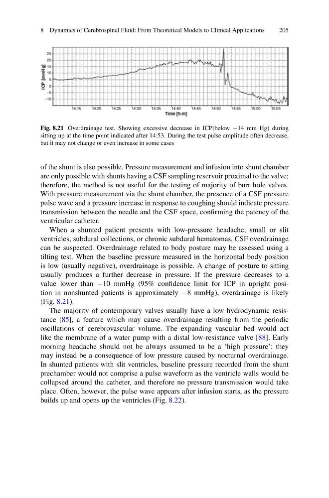
Fig. 8.21 Overdrainage test. Showing excessive decrease in ICP(below −14 mm Hg) during
sitting up at the time point indicated after 14:53. During the test pulse amplitude often decrease,
but it may not change or even increase in some cases
of the shunt is also possible. Pressure measurement and infusion into shunt chamber
are only possible with shunts having a CSF sampling reservoir proximal to the valve;
therefore, the method is not useful for the testing of majority of burr hole valves.
With pressure measurement via the shunt chamber, the presence of a CSF pressure
pulse wave and a pressure increase in response to coughing should indicate pressure
transmission between the needle and the CSF space, confirming the patency of the
ventricular catheter.
When a shunted patient presents with low-pressure headache, small or slit
ventricles, subdural collections, or chronic subdural hematomas, CSF overdrainage
can be suspected. Overdrainage related to body posture may be assessed using a
tilting test. When the baseline pressure measured in the horizontal body position
is low (usually negative), overdrainage is possible. A change of posture to sitting
usually produces a further decrease in pressure. If the pressure decreases to a
value lower than −10 mmHg (95% confidence limit for ICP in upright position in nonshunted patients is approximately −8 mmHg), overdrainage is likely
(Fig. 8.21).
The majority of contemporary valves usually have a low hydrodynamic resistance [85], a feature which may cause overdrainage resulting from the periodic
oscillations of cerebrovascular volume. The expanding vascular bed would act
like the membrane of a water pump with a distal low-resistance valve [88]. Early
morning headache should not be always assumed to be a ‘high pressure’: they
may instead be a consequence of low pressure caused by nocturnal overdrainage.
In shunted patients with slit ventricles, baseline pressure recorded from the shunt
prechamber would not comprise a pulse waveform as the ventricle walls would be
collapsed around the catheter, and therefore no pressure transmission would take
place. Often, however, the pulse wave appears after infusion starts, as the pressure
builds up and opens up the ventricles (Fig. 8.22).
205
Fig. 8.21 Overdrainage test. Showing excessive decrease in ICP(below −14 mm Hg) during
sitting up at the time point indicated after 14:53. During the test pulse amplitude often decrease,
but it may not change or even increase in some cases
of the shunt is also possible. Pressure measurement and infusion into shunt chamber
are only possible with shunts having a CSF sampling reservoir proximal to the valve;
therefore, the method is not useful for the testing of majority of burr hole valves.
With pressure measurement via the shunt chamber, the presence of a CSF pressure
pulse wave and a pressure increase in response to coughing should indicate pressure
transmission between the needle and the CSF space, confirming the patency of the
ventricular catheter.
When a shunted patient presents with low-pressure headache, small or slit
ventricles, subdural collections, or chronic subdural hematomas, CSF overdrainage
can be suspected. Overdrainage related to body posture may be assessed using a
tilting test. When the baseline pressure measured in the horizontal body position
is low (usually negative), overdrainage is possible. A change of posture to sitting
usually produces a further decrease in pressure. If the pressure decreases to a
value lower than −10 mmHg (95% confidence limit for ICP in upright position in nonshunted patients is approximately −8 mmHg), overdrainage is likely
(Fig. 8.21).
The majority of contemporary valves usually have a low hydrodynamic resistance [85], a feature which may cause overdrainage resulting from the periodic
oscillations of cerebrovascular volume. The expanding vascular bed would act
like the membrane of a water pump with a distal low-resistance valve [88]. Early
morning headache should not be always assumed to be a ‘high pressure’: they
may instead be a consequence of low pressure caused by nocturnal overdrainage.
In shunted patients with slit ventricles, baseline pressure recorded from the shunt
prechamber would not comprise a pulse waveform as the ventricle walls would be
collapsed around the catheter, and therefore no pressure transmission would take
place. Often, however, the pulse wave appears after infusion starts, as the pressure
builds up and opens up the ventricles (Fig. 8.22).