

8 Dynamics of Cerebrospinal Fluid: From Theoretical Models to Clinical Applications
197
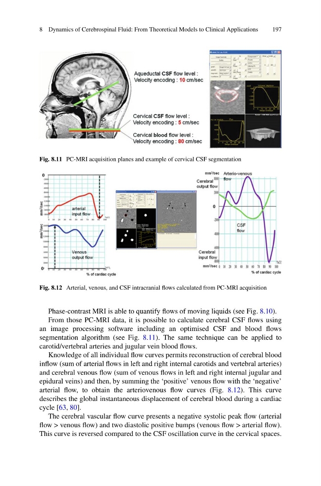
Fig. 8.11 PC-MRI acquisition planes and example of cervical CSF segmentation
Fig. 8.12 Arterial, venous, and CSF intracranial flows calculated from PC-MRI acquisition
Phase-contrast MRI is able to quantify flows of moving liquids (see Fig. 8.10).
From those PC-MRI data, it is possible to calculate cerebral CSF flows using
an image processing software including an optimised CSF and blood flows
segmentation algorithm (see Fig. 8.11). The same technique can be applied to
carotid/vertebral arteries and jugular vein blood flows.
Knowledge of all individual flow curves permits reconstruction of cerebral blood
inflow (sum of arterial flows in left and right internal carotids and vertebral arteries)
and cerebral venous flow (sum of venous flows in left and right internal jugular and
epidural veins) and then, by summing the ‘positive’ venous flow with the ‘negative’
arterial flow, to obtain the arteriovenous flow curves (Fig. 8.12). This curve
describes the global instantaneous displacement of cerebral blood during a cardiac
cycle [63, 80].
The cerebral vascular flow curve presents a negative systolic peak flow (arterial
flow > venous flow) and two diastolic positive bumps (venous flow > arterial flow).
This curve is reversed compared to the CSF oscillation curve in the cervical spaces.
197
Fig. 8.11 PC-MRI acquisition planes and example of cervical CSF segmentation
Fig. 8.12 Arterial, venous, and CSF intracranial flows calculated from PC-MRI acquisition
Phase-contrast MRI is able to quantify flows of moving liquids (see Fig. 8.10).
From those PC-MRI data, it is possible to calculate cerebral CSF flows using
an image processing software including an optimised CSF and blood flows
segmentation algorithm (see Fig. 8.11). The same technique can be applied to
carotid/vertebral arteries and jugular vein blood flows.
Knowledge of all individual flow curves permits reconstruction of cerebral blood
inflow (sum of arterial flows in left and right internal carotids and vertebral arteries)
and cerebral venous flow (sum of venous flows in left and right internal jugular and
epidural veins) and then, by summing the ‘positive’ venous flow with the ‘negative’
arterial flow, to obtain the arteriovenous flow curves (Fig. 8.12). This curve
describes the global instantaneous displacement of cerebral blood during a cardiac
cycle [63, 80].
The cerebral vascular flow curve presents a negative systolic peak flow (arterial
flow > venous flow) and two diastolic positive bumps (venous flow > arterial flow).
This curve is reversed compared to the CSF oscillation curve in the cervical spaces.