

7 Biomechanical Modelling of the Brain for Neuronavigation in Epilepsy Surgery
171
to apply in biomechanical models are uncertain and depend on the situation. In this
case, it was found that the following targets give a mesh of sufficient quality:
• Warpage: 90 ◦
• Length: 0.5 mm
• Jacobian: 0.5
Elements that fail to meet these criteria are isolated into a separate component
and converted into tetrahedral elements before being merged back into the original
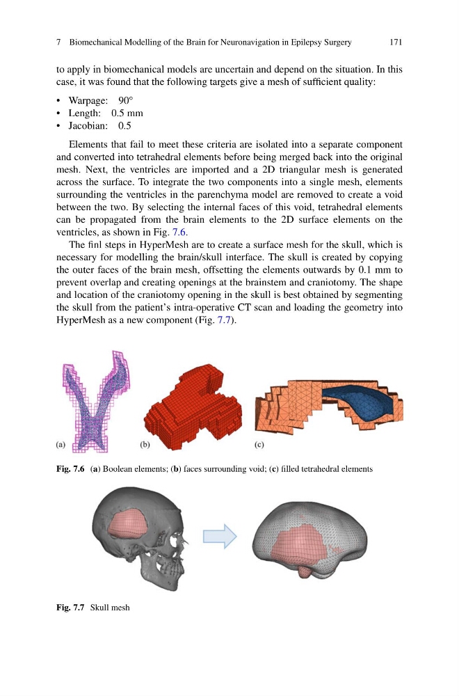
mesh. Next, the ventricles are imported and a 2D triangular mesh is generated
across the surface. To integrate the two components into a single mesh, elements
surrounding the ventricles in the parenchyma model are removed to create a void
between the two. By selecting the internal faces of this void, tetrahedral elements
can be propagated from the brain elements to the 2D surface elements on the
ventricles, as shown in Fig. 7.6.
The finl steps in HyperMesh are to create a surface mesh for the skull, which is
necessary for modelling the brain/skull interface. The skull is created by copying
the outer faces of the brain mesh, offsetting the elements outwards by 0.1 mm to
prevent overlap and creating openings at the brainstem and craniotomy. The shape
and location of the craniotomy opening in the skull is best obtained by segmenting
the skull from the patient’s intra-operative CT scan and loading the geometry into
HyperMesh as a new component (Fig. 7.7).
Fig. 7.6 (a) Boolean elements; (b) faces surrounding void; (c) filled tetrahedral elements
Fig. 7.7 Skull mesh
171
to apply in biomechanical models are uncertain and depend on the situation. In this
case, it was found that the following targets give a mesh of sufficient quality:
• Warpage: 90 ◦
• Length: 0.5 mm
• Jacobian: 0.5
Elements that fail to meet these criteria are isolated into a separate component
and converted into tetrahedral elements before being merged back into the original
mesh. Next, the ventricles are imported and a 2D triangular mesh is generated
across the surface. To integrate the two components into a single mesh, elements
surrounding the ventricles in the parenchyma model are removed to create a void
between the two. By selecting the internal faces of this void, tetrahedral elements
can be propagated from the brain elements to the 2D surface elements on the
ventricles, as shown in Fig. 7.6.
The finl steps in HyperMesh are to create a surface mesh for the skull, which is
necessary for modelling the brain/skull interface. The skull is created by copying
the outer faces of the brain mesh, offsetting the elements outwards by 0.1 mm to
prevent overlap and creating openings at the brainstem and craniotomy. The shape
and location of the craniotomy opening in the skull is best obtained by segmenting
the skull from the patient’s intra-operative CT scan and loading the geometry into
HyperMesh as a new component (Fig. 7.7).
Fig. 7.6 (a) Boolean elements; (b) faces surrounding void; (c) filled tetrahedral elements
Fig. 7.7 Skull mesh