

6 Biomechanical Modelling of the Brain for Neurosurgical Simulation. . .
141
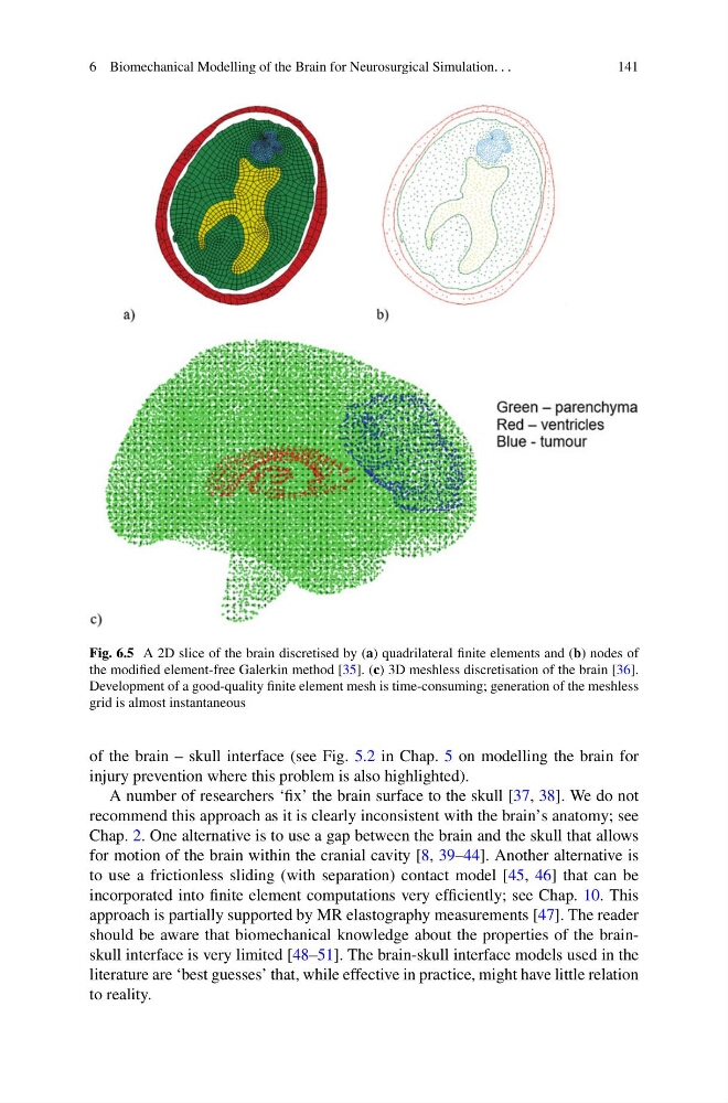
Fig. 6.5 A 2D slice of the brain discretised by (a) quadrilateral finite elements and (b) nodes of
the modified element-free Galerkin method [35]. (c) 3D meshless discretisation of the brain [36].
Development of a good-quality finite element mesh is time-consuming; generation of the meshless
grid is almost instantaneous
of the brain – skull interface (see Fig. 5.2 in Chap. 5 on modelling the brain for
injury prevention where this problem is also highlighted).
A number of researchers ‘fix’ the brain surface to the skull [37, 38]. We do not
recommend this approach as it is clearly inconsistent with the brain’s anatomy; see
Chap. 2. One alternative is to use a gap between the brain and the skull that allows
for motion of the brain within the cranial cavity [8, 39–44]. Another alternative is
to use a frictionless sliding (with separation) contact model [45, 46] that can be
incorporated into finite element computations very efficiently; see Chap. 10. This
approach is partially supported by MR elastography measurements [47]. The reader
should be aware that biomechanical knowledge about the properties of the brainskull interface is very limited [48–51]. The brain-skull interface models used in the
literature are ‘best guesses’ that, while effective in practice, might have little relation
to reality.
141
Fig. 6.5 A 2D slice of the brain discretised by (a) quadrilateral finite elements and (b) nodes of
the modified element-free Galerkin method [35]. (c) 3D meshless discretisation of the brain [36].
Development of a good-quality finite element mesh is time-consuming; generation of the meshless
grid is almost instantaneous
of the brain – skull interface (see Fig. 5.2 in Chap. 5 on modelling the brain for
injury prevention where this problem is also highlighted).
A number of researchers ‘fix’ the brain surface to the skull [37, 38]. We do not
recommend this approach as it is clearly inconsistent with the brain’s anatomy; see
Chap. 2. One alternative is to use a gap between the brain and the skull that allows
for motion of the brain within the cranial cavity [8, 39–44]. Another alternative is
to use a frictionless sliding (with separation) contact model [45, 46] that can be
incorporated into finite element computations very efficiently; see Chap. 10. This
approach is partially supported by MR elastography measurements [47]. The reader
should be aware that biomechanical knowledge about the properties of the brainskull interface is very limited [48–51]. The brain-skull interface models used in the
literature are ‘best guesses’ that, while effective in practice, might have little relation
to reality.