

5 Modelling of the Brain for Injury Simulation and Prevention
111
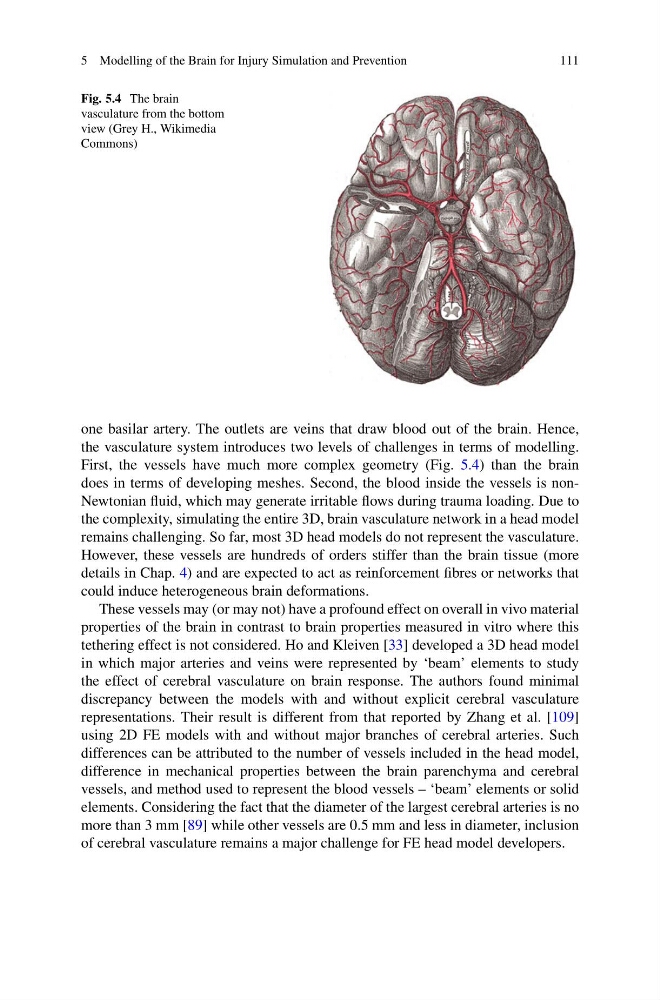
Fig. 5.4 The brain
vasculature from the bottom
view (Grey H., Wikimedia
Commons)
one basilar artery. The outlets are veins that draw blood out of the brain. Hence,
the vasculature system introduces two levels of challenges in terms of modelling.
First, the vessels have much more complex geometry (Fig. 5.4) than the brain
does in terms of developing meshes. Second, the blood inside the vessels is nonNewtonian fluid, which may generate irritable flows during trauma loading. Due to
the complexity, simulating the entire 3D, brain vasculature network in a head model
remains challenging. So far, most 3D head models do not represent the vasculature.
However, these vessels are hundreds of orders stiffer than the brain tissue (more
details in Chap. 4) and are expected to act as reinforcement fibres or networks that
could induce heterogeneous brain deformations.
These vessels may (or may not) have a profound effect on overall in vivo material
properties of the brain in contrast to brain properties measured in vitro where this
tethering effect is not considered. Ho and Kleiven [33] developed a 3D head model
in which major arteries and veins were represented by ‘beam’ elements to study
the effect of cerebral vasculature on brain response. The authors found minimal
discrepancy between the models with and without explicit cerebral vasculature
representations. Their result is different from that reported by Zhang et al. [109]
using 2D FE models with and without major branches of cerebral arteries. Such
differences can be attributed to the number of vessels included in the head model,
difference in mechanical properties between the brain parenchyma and cerebral
vessels, and method used to represent the blood vessels – ‘beam’ elements or solid
elements. Considering the fact that the diameter of the largest cerebral arteries is no
more than 3 mm [89] while other vessels are 0.5 mm and less in diameter, inclusion
of cerebral vasculature remains a major challenge for FE head model developers.
111
Fig. 5.4 The brain
vasculature from the bottom
view (Grey H., Wikimedia
Commons)
one basilar artery. The outlets are veins that draw blood out of the brain. Hence,
the vasculature system introduces two levels of challenges in terms of modelling.
First, the vessels have much more complex geometry (Fig. 5.4) than the brain
does in terms of developing meshes. Second, the blood inside the vessels is nonNewtonian fluid, which may generate irritable flows during trauma loading. Due to
the complexity, simulating the entire 3D, brain vasculature network in a head model
remains challenging. So far, most 3D head models do not represent the vasculature.
However, these vessels are hundreds of orders stiffer than the brain tissue (more
details in Chap. 4) and are expected to act as reinforcement fibres or networks that
could induce heterogeneous brain deformations.
These vessels may (or may not) have a profound effect on overall in vivo material
properties of the brain in contrast to brain properties measured in vitro where this
tethering effect is not considered. Ho and Kleiven [33] developed a 3D head model
in which major arteries and veins were represented by ‘beam’ elements to study
the effect of cerebral vasculature on brain response. The authors found minimal
discrepancy between the models with and without explicit cerebral vasculature
representations. Their result is different from that reported by Zhang et al. [109]
using 2D FE models with and without major branches of cerebral arteries. Such
differences can be attributed to the number of vessels included in the head model,
difference in mechanical properties between the brain parenchyma and cerebral
vessels, and method used to represent the blood vessels – ‘beam’ elements or solid
elements. Considering the fact that the diameter of the largest cerebral arteries is no
more than 3 mm [89] while other vessels are 0.5 mm and less in diameter, inclusion
of cerebral vasculature remains a major challenge for FE head model developers.