

110
K. H. Yang and H. Mao
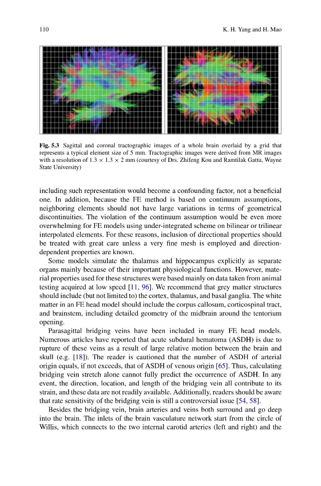
Fig. 5.3 Sagittal and coronal tractographic images of a whole brain overlaid by a grid that
represents a typical element size of 5 mm. Tractographic images were derived from MR images
with a resolution of 1.3 × 1.3 × 2 mm (courtesy of Drs. Zhifeng Kou and Ramtilak Gattu, Wayne
State University)
including such representation would become a confounding factor, not a beneficial
one. In addition, because the FE method is based on continuum assumptions,
neighboring elements should not have large variations in terms of geometrical
discontinuities. The violation of the continuum assumption would be even more
overwhelming for FE models using under-integrated scheme on bilinear or trilinear
interpolated elements. For these reasons, inclusion of directional properties should
be treated with great care unless a very fine mesh is employed and directiondependent properties are known.
Some models simulate the thalamus and hippocampus explicitly as separate
organs mainly because of their important physiological functions. However, material properties used for these structures were based mainly on data taken from animal
testing acquired at low speed [11, 96]. We recommend that grey matter structures
should include (but not limited to) the cortex, thalamus, and basal ganglia. The white
matter in an FE head model should include the corpus callosum, corticospinal tract,
and brainstem, including detailed geometry of the midbrain around the tentorium
opening.
Parasagittal bridging veins have been included in many FE head models.
Numerous articles have reported that acute subdural hematoma (ASDH) is due to
rupture of these veins as a result of large relative motion between the brain and
skull (e.g. [18]). The reader is cautioned that the number of ASDH of arterial
origin equals, if not exceeds, that of ASDH of venous origin [65]. Thus, calculating
bridging vein stretch alone cannot fully predict the occurrence of ASDH. In any
event, the direction, location, and length of the bridging vein all contribute to its
strain, and these data are not readily available. Additionally, readers should be aware
that rate sensitivity of the bridging vein is still a controversial issue [54, 58].
Besides the bridging vein, brain arteries and veins both surround and go deep
into the brain. The inlets of the brain vasculature network start from the circle of
Willis, which connects to the two internal carotid arteries (left and right) and the
K. H. Yang and H. Mao
Fig. 5.3 Sagittal and coronal tractographic images of a whole brain overlaid by a grid that
represents a typical element size of 5 mm. Tractographic images were derived from MR images
with a resolution of 1.3 × 1.3 × 2 mm (courtesy of Drs. Zhifeng Kou and Ramtilak Gattu, Wayne
State University)
including such representation would become a confounding factor, not a beneficial
one. In addition, because the FE method is based on continuum assumptions,
neighboring elements should not have large variations in terms of geometrical
discontinuities. The violation of the continuum assumption would be even more
overwhelming for FE models using under-integrated scheme on bilinear or trilinear
interpolated elements. For these reasons, inclusion of directional properties should
be treated with great care unless a very fine mesh is employed and directiondependent properties are known.
Some models simulate the thalamus and hippocampus explicitly as separate
organs mainly because of their important physiological functions. However, material properties used for these structures were based mainly on data taken from animal
testing acquired at low speed [11, 96]. We recommend that grey matter structures
should include (but not limited to) the cortex, thalamus, and basal ganglia. The white
matter in an FE head model should include the corpus callosum, corticospinal tract,
and brainstem, including detailed geometry of the midbrain around the tentorium
opening.
Parasagittal bridging veins have been included in many FE head models.
Numerous articles have reported that acute subdural hematoma (ASDH) is due to
rupture of these veins as a result of large relative motion between the brain and
skull (e.g. [18]). The reader is cautioned that the number of ASDH of arterial
origin equals, if not exceeds, that of ASDH of venous origin [65]. Thus, calculating
bridging vein stretch alone cannot fully predict the occurrence of ASDH. In any
event, the direction, location, and length of the bridging vein all contribute to its
strain, and these data are not readily available. Additionally, readers should be aware
that rate sensitivity of the bridging vein is still a controversial issue [54, 58].
Besides the bridging vein, brain arteries and veins both surround and go deep
into the brain. The inlets of the brain vasculature network start from the circle of
Willis, which connects to the two internal carotid arteries (left and right) and the