

5 EEG Source Imaging and Multimodal Neuroimaging
113
x i
x
F
i x
E
i
and s i [s
F
i s
E
i ]
(5.48)
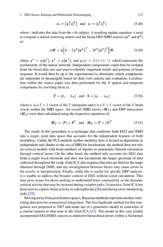
where i indicates the data from the i-th subject. A resulting update equation is used
to compute a shared unmixing matrix and the fused ERP fMRI sources (u
F and u
E )
as:
W η
I − 2 y
E
u
E
T − 2I
F
u
F
T
W
(5.49)
where y
E
g
u
E
, y
F
g
u
F
, and g(x) 1/(1 + e
−x ), which represents the
nonlinearity of the neural network. Independent components could then be isolated
from the fused data sets and used to identify important motifs and patterns of brain
response. It would then be up to the experimenter to determine which components
are important or meaningful based on their own criteria and evaluation. Localization within the source paper was then performed for the N spatial and temporal
components by rewriting them as:
T [t 1 . . . t N ] and S [s 1 . . . s N ]
(5.50)
where t i is a T × 1 vector of the T timepoints and s is a V × 1 vector of the V brain
voxels within the MRI space. An overall fMRI movie (M F ) and ERP timecourse
(M E ) were then calculated using the respective equations of:
M F |T | × S
T and M E T × |S|
T
(5.51)
The result of this procedure is a technique that combines both EEG and fMRI
into a single, joint data space that accounts for the independent features of both
modalities. Unlike the PLS method, neither modality here is treated as dependent or
independent and, thanks to the use of MRI for localization, the method does not rely
on cortical models with fixed numbers of dipoles or potentially blurred calculation
through cortical layers. On the other hand, the method only accounts for EEG data
from a single focal electrode and does not incorporate the larger spectrum of data
collected throughout the scalp. Joint ICA also requires that data are fitted to the maps
obtained through fMRI, and any misalignment between theses may cause errors in
the results or interpretation. Finally, while this is useful for specific ERP analysis,
it is unable to address the broader context of EEG without serial calculation. This
may pose issues for those seeking to understand brain connectivity or the dynamic
cortical activity that may be incurred during complex tasks. In practice, Joint ICA has
been used to explore brain activity in schizophrenia [26] and during error-monitoring
tasks [22].
Moving away from joint feature spaces, Bayesian methods represent another interesting direction for symmetrical integration. The first landmark method for this integration was proposed in 2007 and made use of a generative model in somewhat of
a similar manner to that seen in the Joint ICA [21]. The model in this case jointly
incorporated EEG/fMRI sources as unknown hierarchical priors within a Variatonal
113
x i
x
F
i x
E
i
and s i [s
F
i s
E
i ]
(5.48)
where i indicates the data from the i-th subject. A resulting update equation is used
to compute a shared unmixing matrix and the fused ERP fMRI sources (u
F and u
E )
as:
W η
I − 2 y
E
u
E
T − 2I
F
u
F
T
W
(5.49)
where y
E
g
u
E
, y
F
g
u
F
, and g(x) 1/(1 + e
−x ), which represents the
nonlinearity of the neural network. Independent components could then be isolated
from the fused data sets and used to identify important motifs and patterns of brain
response. It would then be up to the experimenter to determine which components
are important or meaningful based on their own criteria and evaluation. Localization within the source paper was then performed for the N spatial and temporal
components by rewriting them as:
T [t 1 . . . t N ] and S [s 1 . . . s N ]
(5.50)
where t i is a T × 1 vector of the T timepoints and s is a V × 1 vector of the V brain
voxels within the MRI space. An overall fMRI movie (M F ) and ERP timecourse
(M E ) were then calculated using the respective equations of:
M F |T | × S
T and M E T × |S|
T
(5.51)
The result of this procedure is a technique that combines both EEG and fMRI
into a single, joint data space that accounts for the independent features of both
modalities. Unlike the PLS method, neither modality here is treated as dependent or
independent and, thanks to the use of MRI for localization, the method does not rely
on cortical models with fixed numbers of dipoles or potentially blurred calculation
through cortical layers. On the other hand, the method only accounts for EEG data
from a single focal electrode and does not incorporate the larger spectrum of data
collected throughout the scalp. Joint ICA also requires that data are fitted to the maps
obtained through fMRI, and any misalignment between theses may cause errors in
the results or interpretation. Finally, while this is useful for specific ERP analysis,
it is unable to address the broader context of EEG without serial calculation. This
may pose issues for those seeking to understand brain connectivity or the dynamic
cortical activity that may be incurred during complex tasks. In practice, Joint ICA has
been used to explore brain activity in schizophrenia [26] and during error-monitoring
tasks [22].
Moving away from joint feature spaces, Bayesian methods represent another interesting direction for symmetrical integration. The first landmark method for this integration was proposed in 2007 and made use of a generative model in somewhat of
a similar manner to that seen in the Joint ICA [21]. The model in this case jointly
incorporated EEG/fMRI sources as unknown hierarchical priors within a Variatonal