

114
Y. Zhang
Bayesian learning scheme, resulting in an estimate of the common spatial profile
for the modalities. Included as a part of this approach is an estimation of the spatial structure of the EEG/fMRI coupling and uncoupling. As with EEG-only data,
probabilistic approaches are of great interest because they are robust to imprecision
and accommodate a degree of error without greatly inhibiting localization ability.
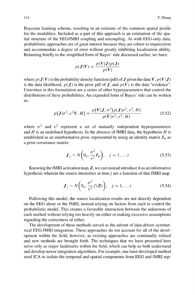
Returning briefly to the simplified form of Bayes’ rule discussed earlier, we have:
p( J|V )
p(V | J) p( J)
p(V )
where p( J|V ) is the probability density function (pdf) of J given the data V, p(V | J)
is the data likelihood, p( J) is the prior pdf of J, and p(V ) is the data “evidence”.
Unwritten in this formulation are a series of other hyperparameters that control the
distributions of these probabilities. An expanded form of Bayes’ rule can be written
as:
p
J|σ
2
, ,
2 V , H
p
V | J, σ
2
p( J|σ
2
, ,
2
, H )
p(V |σ 2 , , 2 , H )
(5.52)
where σ
2 and
2 represent a set of mutually independent hyperparameters
and H is an undefined hypothesis. In the absence of fMRI data, the hypothesis H is
established as an uninformative prior, represented by using an identity matrix I n as
a prior covariance matrix:
J j ∼ N
0 n ,
σ
2
2 I n
, j 1, . . . t
(5.53)
Knowing the fMRI activation map, Z, we can instead introduce it as an informative
hypothesis wherein the source intensities at time j are a function of that fMRI map.
J j ∼ N
0 n ,
σ
2
2 f (Z)
, j 1, . . . t
(5.54)
Following this model, the source localization results are not directly dependent
on the EEG alone or the fMRI, instead relying on factors from each to control the
probabilistic model. This creates a favorable interaction between the unknowns of
each method without relying too heavily on either or making excessive assumptions
regarding the correctness of either.
The development of these methods served as the advent of data-driven symmetrical EEG-fMRI integration. These approaches do not account for all of the development within the field; however, as existing approaches are continually refined
and new methods are brought forth. The techniques that we have presented here
serve only as major landmarks within the field, which can help us both understand
and develop newer integration algorithms. For example, one later-developed method
used ICA to isolate the temporal and spatial components from EEG and fMRI sep-
Y. Zhang
Bayesian learning scheme, resulting in an estimate of the common spatial profile
for the modalities. Included as a part of this approach is an estimation of the spatial structure of the EEG/fMRI coupling and uncoupling. As with EEG-only data,
probabilistic approaches are of great interest because they are robust to imprecision
and accommodate a degree of error without greatly inhibiting localization ability.
Returning briefly to the simplified form of Bayes’ rule discussed earlier, we have:
p( J|V )
p(V | J) p( J)
p(V )
where p( J|V ) is the probability density function (pdf) of J given the data V, p(V | J)
is the data likelihood, p( J) is the prior pdf of J, and p(V ) is the data “evidence”.
Unwritten in this formulation are a series of other hyperparameters that control the
distributions of these probabilities. An expanded form of Bayes’ rule can be written
as:
p
J|σ
2
, ,
2 V , H
p
V | J, σ
2
p( J|σ
2
, ,
2
, H )
p(V |σ 2 , , 2 , H )
(5.52)
where σ
2 and
2 represent a set of mutually independent hyperparameters
and H is an undefined hypothesis. In the absence of fMRI data, the hypothesis H is
established as an uninformative prior, represented by using an identity matrix I n as
a prior covariance matrix:
J j ∼ N
0 n ,
σ
2
2 I n
, j 1, . . . t
(5.53)
Knowing the fMRI activation map, Z, we can instead introduce it as an informative
hypothesis wherein the source intensities at time j are a function of that fMRI map.
J j ∼ N
0 n ,
σ
2
2 f (Z)
, j 1, . . . t
(5.54)
Following this model, the source localization results are not directly dependent
on the EEG alone or the fMRI, instead relying on factors from each to control the
probabilistic model. This creates a favorable interaction between the unknowns of
each method without relying too heavily on either or making excessive assumptions
regarding the correctness of either.
The development of these methods served as the advent of data-driven symmetrical EEG-fMRI integration. These approaches do not account for all of the development within the field; however, as existing approaches are continually refined
and new methods are brought forth. The techniques that we have presented here
serve only as major landmarks within the field, which can help us both understand
and develop newer integration algorithms. For example, one later-developed method
used ICA to isolate the temporal and spatial components from EEG and fMRI sep-