

3.5 Conclusion
Numerous radiolabeled porphyrins and phthalocyanines and their radiopharmaceutical applications such as biodistribution, tumor uptake, PET/SPECT imaging
and PDT have been summarized in this chapter. Tumor accumulation depends on
many factors including the characteristics of the tumor, the porphyrin or PC, the
type of targeting groups and the choice of radioisotopes. Many radiolabeled porphyrins exhibit high tumor uptake that hold potential to be translated into imaging
or therapeutic agents for cancer applications.
References
1. M. Biesaga, K. Pyrzyńska, M. Trojanowicz, Porphyrins in analytical chemistry. A review.
Talanta 51(2), 209–224 (2000)
2. H. Huang, W. Song, J. Rieffel, J.F. Lovell, Emerging applications of porphyrins in
photomedicine. Front Phys. 3, 23 (2015)
3. Y. Zhang, J.F. Lovell, Porphyrins as theranostic agents from prehistoric to modern times.
Theranostics 2(9), 905–915 (2012)
4. D. Kessel, Hematoporphyrin and HPD: photophysics, photochemistry and phototherapy.
Photochem. Photobiol. 39(s1), 851–859 (1984)
5. K.M. Smith, K.M. Kadish, R. Guilard, Handbook of Porphyrin Science: With Applications to
Chemistry, Physics, Materials Science, Engineering, Biology and Medicine, vol 3, (World
Scientific, 2012)
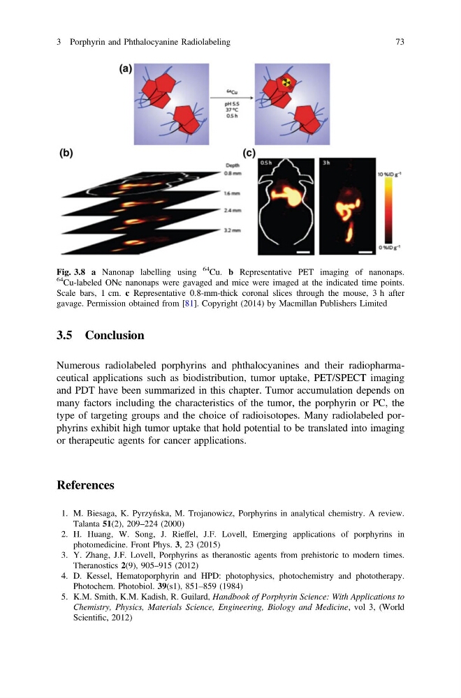
Fig. 3.8 a Nanonap labelling using
64
Cu. b Representative PET imaging of nanonaps.
64
Cu-labeled ONc nanonaps were gavaged and mice were imaged at the indicated time points.
Scale bars, 1 cm. c Representative 0.8-mm-thick coronal slices through the mouse, 3 h after
gavage. Permission obtained from [81]. Copyright (2014) by Macmillan Publishers Limited
3 Porphyrin and Phthalocyanine Radiolabeling
73
Numerous radiolabeled porphyrins and phthalocyanines and their radiopharmaceutical applications such as biodistribution, tumor uptake, PET/SPECT imaging
and PDT have been summarized in this chapter. Tumor accumulation depends on
many factors including the characteristics of the tumor, the porphyrin or PC, the
type of targeting groups and the choice of radioisotopes. Many radiolabeled porphyrins exhibit high tumor uptake that hold potential to be translated into imaging
or therapeutic agents for cancer applications.
References
1. M. Biesaga, K. Pyrzyńska, M. Trojanowicz, Porphyrins in analytical chemistry. A review.
Talanta 51(2), 209–224 (2000)
2. H. Huang, W. Song, J. Rieffel, J.F. Lovell, Emerging applications of porphyrins in
photomedicine. Front Phys. 3, 23 (2015)
3. Y. Zhang, J.F. Lovell, Porphyrins as theranostic agents from prehistoric to modern times.
Theranostics 2(9), 905–915 (2012)
4. D. Kessel, Hematoporphyrin and HPD: photophysics, photochemistry and phototherapy.
Photochem. Photobiol. 39(s1), 851–859 (1984)
5. K.M. Smith, K.M. Kadish, R. Guilard, Handbook of Porphyrin Science: With Applications to
Chemistry, Physics, Materials Science, Engineering, Biology and Medicine, vol 3, (World
Scientific, 2012)
Fig. 3.8 a Nanonap labelling using
64
Cu. b Representative PET imaging of nanonaps.
64
Cu-labeled ONc nanonaps were gavaged and mice were imaged at the indicated time points.
Scale bars, 1 cm. c Representative 0.8-mm-thick coronal slices through the mouse, 3 h after
gavage. Permission obtained from [81]. Copyright (2014) by Macmillan Publishers Limited
3 Porphyrin and Phthalocyanine Radiolabeling
73