

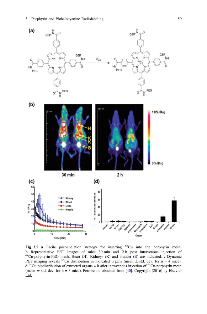
Fig. 3.5 a Facile post-chelation strategy for inserting
64
Cu into the porphyrin mesh.
b Representative PET images of mice 30 min and 2 h post intravenous injection of
64
Cu-porphyrin-PEG mesh. Heart (H), Kidneys (K) and bladder (B) are indicated. c Dynamic
PET imaging reveals
64
Cu distribution in indicated organs (mean ± std. dev. for n = 4 mice).
d
64
Cu biodistribution of extracted organs 4 h after intravenous injection of
64
Cu-porphyrin mesh
(mean ± std. dev. for n = 3 mice). Permission obtained from [40]. Copyright (2016) by Elsevier
Ltd.
3 Porphyrin and Phthalocyanine Radiolabeling
59
64
Cu into the porphyrin mesh.
b Representative PET images of mice 30 min and 2 h post intravenous injection of
64
Cu-porphyrin-PEG mesh. Heart (H), Kidneys (K) and bladder (B) are indicated. c Dynamic
PET imaging reveals
64
Cu distribution in indicated organs (mean ± std. dev. for n = 4 mice).
d
64
Cu biodistribution of extracted organs 4 h after intravenous injection of
64
Cu-porphyrin mesh
(mean ± std. dev. for n = 3 mice). Permission obtained from [40]. Copyright (2016) by Elsevier
Ltd.
3 Porphyrin and Phthalocyanine Radiolabeling
59