

22.5 Multimodal Molecular Imaging
The multimodal probes development has been progressing rapidly due to
advancements in hybrid imaging. Radiolabeled NPs have many strengths as multimodal probes, for example, the injection of a single agent for multimodal imaging.
In addition, the signal is constant at the target region and no difference is found in
biodistribution, which may arise when more than two agents are utilized.
Among several molecular imaging modalities, single modality is not enough for
obtaining all the needed information [5]. For example, fluorescence signal quantification is difficult in vivo, especially in deep tissues. MRI has good resolution but
relatively low sensitivity, whereas nuclear imaging has good sensitivity but comparatively low resolution. A combined utilization of several imaging modalities can
offer synergistic benefits. Combination of optical imaging with three-dimensional
tomography, such as PET, SPECT, or MRI, can facilitate noninvasive imaging
in vivo with higher diagnostic value. NPs have large surface areas where multiple
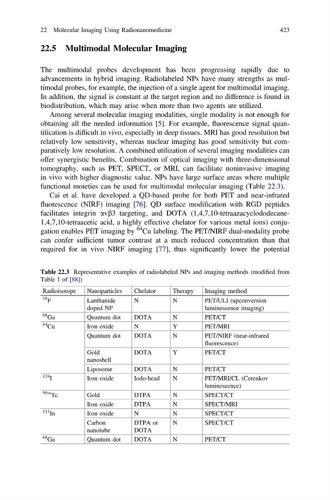
functional moieties can be used for multimodal molecular imaging (Table 22.3).
Cai et al. have developed a QD-based probe for both PET and near-infrared
fluorescence (NIRF) imaging [76]. QD surface modification with RGD peptides
facilitates integrin avb3 targeting, and DOTA (1,4,7,10-tetraazacyclododecane1,4,7,10-tetraacetic acid, a highly effective chelator for various metal ions) conjugation enables PET imaging by
64 Cu labeling. The PET/NIRF dual-modality probe
can confer sufficient tumor contrast at a much reduced concentration than that
required for in vivo NIRF imaging [77], thus significantly lower the potential
Table 22.3 Representative examples of radiolabeled NPs and imaging methods (modified from
Table 1 of [88])
Radioisotope
Nanoparticles
Chelator
Therapy
Imaging method
18
F
Lanthanide
doped NP
N
N
PET/ULI (upconversion
luminescence imaging)
68
Ga
Quantum dot
DOTA
N
PET/CT
64
Cu
Iron oxide
N
Y
PET/MRI
Quantum dot
DOTA
N
PET/NIRF (near-infrared
fluorescence)
Gold
nanoshell
DOTA
Y
PET/CT
Liposome
DOTA
N
PET/CT
124
I
Iron oxide
Iodo-bead
N
PET/MRI/CL (Cerenkov
luminescence)
99m
Tc
Gold
DTPA
N
SPECT/CT
Iron oxide
DTPA
N
SPECT/MRI
111
In
Iron oxide
N
N
SPECT/CT
Carbon
nanotube
DTPA or
DOTA
N
SPECT/CT
68
Ga
Quantum dot
DOTA
N
PET/CT
22 Molecular Imaging Using Radionanomedicine
423
The multimodal probes development has been progressing rapidly due to
advancements in hybrid imaging. Radiolabeled NPs have many strengths as multimodal probes, for example, the injection of a single agent for multimodal imaging.
In addition, the signal is constant at the target region and no difference is found in
biodistribution, which may arise when more than two agents are utilized.
Among several molecular imaging modalities, single modality is not enough for
obtaining all the needed information [5]. For example, fluorescence signal quantification is difficult in vivo, especially in deep tissues. MRI has good resolution but
relatively low sensitivity, whereas nuclear imaging has good sensitivity but comparatively low resolution. A combined utilization of several imaging modalities can
offer synergistic benefits. Combination of optical imaging with three-dimensional
tomography, such as PET, SPECT, or MRI, can facilitate noninvasive imaging
in vivo with higher diagnostic value. NPs have large surface areas where multiple
functional moieties can be used for multimodal molecular imaging (Table 22.3).
Cai et al. have developed a QD-based probe for both PET and near-infrared
fluorescence (NIRF) imaging [76]. QD surface modification with RGD peptides
facilitates integrin avb3 targeting, and DOTA (1,4,7,10-tetraazacyclododecane1,4,7,10-tetraacetic acid, a highly effective chelator for various metal ions) conjugation enables PET imaging by
64 Cu labeling. The PET/NIRF dual-modality probe
can confer sufficient tumor contrast at a much reduced concentration than that
required for in vivo NIRF imaging [77], thus significantly lower the potential
Table 22.3 Representative examples of radiolabeled NPs and imaging methods (modified from
Table 1 of [88])
Radioisotope
Nanoparticles
Chelator
Therapy
Imaging method
18
F
Lanthanide
doped NP
N
N
PET/ULI (upconversion
luminescence imaging)
68
Ga
Quantum dot
DOTA
N
PET/CT
64
Cu
Iron oxide
N
Y
PET/MRI
Quantum dot
DOTA
N
PET/NIRF (near-infrared
fluorescence)
Gold
nanoshell
DOTA
Y
PET/CT
Liposome
DOTA
N
PET/CT
124
I
Iron oxide
Iodo-bead
N
PET/MRI/CL (Cerenkov
luminescence)
99m
Tc
Gold
DTPA
N
SPECT/CT
Iron oxide
DTPA
N
SPECT/MRI
111
In
Iron oxide
N
N
SPECT/CT
Carbon
nanotube
DTPA or
DOTA
N
SPECT/CT
68
Ga
Quantum dot
DOTA
N
PET/CT
22 Molecular Imaging Using Radionanomedicine
423