

Typically,
11 C (t1/2 = 20 min) is not commonly utilized with NPs due to its
short half-life. Nevertheless, a study showed its possibility.
11 C-methyl iodide was
used as a methylation agent to react with carboxylic acid and amine functional
groups bound to NPs, which was finally used as a PET/MRI imaging agent [71].
Similar,
13
N (t1/2 = 9.97 min) requires the direct bombardment of Al 2 O 3 NPs, thus
nullify a radiolabeling step for in vivo usage and allow imaging until 68 min after
injection [72]. The isotope
68 Ga (t1/2 = 68 min) has a half-life that is sufficient for
the in vivo imaging of small and short-circulating NPs. PEGylated IONPs was used
for SLN imaging, thus allowing PET/MRI/Cherenkov luminescence imaging
(CLI) [73].
18 F (t1/2 = 110 min) is attached to NPs via synthetic methods and
imaging is feasible until 8 h after injection. A substitution to synthetic methods is
the usage of a proton beam to bombard TiO 2 NPs, providing
18 F-intrinsically
radiolabeled NP.
If a longer in vivo time is needed,
64 Cu (t1/2 = 12.7 h) permits imaging until
48 h after injection.
64 Cu can be included into porphysomes until 48 h after
injection. Most importantly, radiolabeled porphysomes can retain their size and
optical properties, and have a nearly same biological properties of cold liposomes,
thus demonstrating that radiolabeling does not alter the characteristics of the particles [74]. The PET isotopes with the longest half-lives utilized for NP imaging are
89 Zr (t1/2 = 78.4 h) and
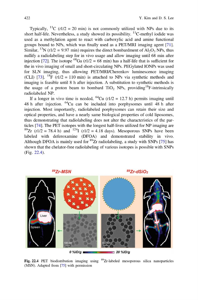
124 I (t1/2 = 4.18 days). Mesoporous SNPs have been
labeled with deferoxamine (DFOA) and demonstrated stability in vivo.
Although DFOA is mainly used for
89
Zr radiolabeling, a study with SNPs [75] has
shown that the chelator-free radiolabeling of various isotopes is possible with SNPs
(Fig. 22.4).
Fig. 22.4 PET biodistribution imaging using
89
Zr-labeled mesoporous silica nanoparticles
(MSN). Adapted from [75] with permission
422
Y. Kim and D. S. Lee
11 C (t1/2 = 20 min) is not commonly utilized with NPs due to its
short half-life. Nevertheless, a study showed its possibility.
11 C-methyl iodide was
used as a methylation agent to react with carboxylic acid and amine functional
groups bound to NPs, which was finally used as a PET/MRI imaging agent [71].
Similar,
13
N (t1/2 = 9.97 min) requires the direct bombardment of Al 2 O 3 NPs, thus
nullify a radiolabeling step for in vivo usage and allow imaging until 68 min after
injection [72]. The isotope
68 Ga (t1/2 = 68 min) has a half-life that is sufficient for
the in vivo imaging of small and short-circulating NPs. PEGylated IONPs was used
for SLN imaging, thus allowing PET/MRI/Cherenkov luminescence imaging
(CLI) [73].
18 F (t1/2 = 110 min) is attached to NPs via synthetic methods and
imaging is feasible until 8 h after injection. A substitution to synthetic methods is
the usage of a proton beam to bombard TiO 2 NPs, providing
18 F-intrinsically
radiolabeled NP.
If a longer in vivo time is needed,
64 Cu (t1/2 = 12.7 h) permits imaging until
48 h after injection.
64 Cu can be included into porphysomes until 48 h after
injection. Most importantly, radiolabeled porphysomes can retain their size and
optical properties, and have a nearly same biological properties of cold liposomes,
thus demonstrating that radiolabeling does not alter the characteristics of the particles [74]. The PET isotopes with the longest half-lives utilized for NP imaging are
89 Zr (t1/2 = 78.4 h) and
124 I (t1/2 = 4.18 days). Mesoporous SNPs have been
labeled with deferoxamine (DFOA) and demonstrated stability in vivo.
Although DFOA is mainly used for
89
Zr radiolabeling, a study with SNPs [75] has
shown that the chelator-free radiolabeling of various isotopes is possible with SNPs
(Fig. 22.4).
Fig. 22.4 PET biodistribution imaging using
89
Zr-labeled mesoporous silica nanoparticles
(MSN). Adapted from [75] with permission
422
Y. Kim and D. S. Lee