

22.1 Molecular Imaging by Nanotechnology
Nanotechnology has great potential for the detection and diagnosis of cancers and
nanoparticles (NPs) can be used in the personalized treatment of various diseases
[1]. In general, the diameter of a NP is between one to hundreds nanometers. NPs
can interact with biomolecules both on the surface of and inside cells, which could
be helpful in the diagnosis and treatment of disease. One major application of
nanotechnology is in medicine [2].
Molecular imaging is defined as in vivo characterization and measurement of
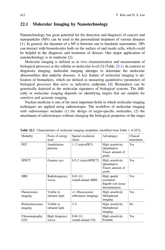
biological processes at the cellular or molecular level [3] (Table 22.1). In contrast to
diagnostic imaging, molecular imaging attempts to determine the molecular
abnormalities that underlie diseases. A key feature of molecular imaging is utilization of biomarkers, which are defined as measuring quantitative parameters of
biological processes that serve as indicative endpoints [4]. Biomarkers can be
generically depicted as the molecular signatures of biological systems. The difficulty in molecular imaging depends on identifying targets that are suitable for
sensitive and accurate imaging.
Nuclear medicine is one of the most important fields in which molecular imaging
techniques are applied using radioisotopes. The workflow of molecular imaging
with radioisotopes includes (1) the design of target-specific molecules, (2) the
attachment of radioisotopes without changing the biological properties of the target
Table 22.1 Characteristics of molecular imaging modalities (modified from Table 1 of [87])
Modality
Form of energy
used
Spatial resolution
(mm)
Advantages
Clinical
translation
PET
Annihilation
photons
1–2 (microPET)
High sensitivity
Quantitative
Tracer amount of
probe
Yes
SPECT
Gamma rays
0.5–2 (microSPECT) High sensitivity
Quantitative
Tracer amount of
probe
Yes
MRI
Radiofrequency
waves
0.01–0.1
(small-animal MRI)
High spatial
resolution
Superb soft tissue
discrimination
Yes
Fluorescence
imaging
Visible to
infrared light
<1 (fluorescence
reflectance imaging)
High sensitivity
Multiplexed
imaging
Yes
Bioluminescence
imaging
Visible to
infrared light
3–5
High sensitivity
Multiplexed
imaging
No
Ultrasonography
(US)
High frequency
waves
0.04–0.1
(small-animal US)
High sensitivity
Portable
Yes
412
Y. Kim and D. S. Lee
Nanotechnology has great potential for the detection and diagnosis of cancers and
nanoparticles (NPs) can be used in the personalized treatment of various diseases
[1]. In general, the diameter of a NP is between one to hundreds nanometers. NPs
can interact with biomolecules both on the surface of and inside cells, which could
be helpful in the diagnosis and treatment of disease. One major application of
nanotechnology is in medicine [2].
Molecular imaging is defined as in vivo characterization and measurement of
biological processes at the cellular or molecular level [3] (Table 22.1). In contrast to
diagnostic imaging, molecular imaging attempts to determine the molecular
abnormalities that underlie diseases. A key feature of molecular imaging is utilization of biomarkers, which are defined as measuring quantitative parameters of
biological processes that serve as indicative endpoints [4]. Biomarkers can be
generically depicted as the molecular signatures of biological systems. The difficulty in molecular imaging depends on identifying targets that are suitable for
sensitive and accurate imaging.
Nuclear medicine is one of the most important fields in which molecular imaging
techniques are applied using radioisotopes. The workflow of molecular imaging
with radioisotopes includes (1) the design of target-specific molecules, (2) the
attachment of radioisotopes without changing the biological properties of the target
Table 22.1 Characteristics of molecular imaging modalities (modified from Table 1 of [87])
Modality
Form of energy
used
Spatial resolution
(mm)
Advantages
Clinical
translation
PET
Annihilation
photons
1–2 (microPET)
High sensitivity
Quantitative
Tracer amount of
probe
Yes
SPECT
Gamma rays
0.5–2 (microSPECT) High sensitivity
Quantitative
Tracer amount of
probe
Yes
MRI
Radiofrequency
waves
0.01–0.1
(small-animal MRI)
High spatial
resolution
Superb soft tissue
discrimination
Yes
Fluorescence
imaging
Visible to
infrared light
<1 (fluorescence
reflectance imaging)
High sensitivity
Multiplexed
imaging
Yes
Bioluminescence
imaging
Visible to
infrared light
3–5
High sensitivity
Multiplexed
imaging
No
Ultrasonography
(US)
High frequency
waves
0.04–0.1
(small-animal US)
High sensitivity
Portable
Yes
412
Y. Kim and D. S. Lee