

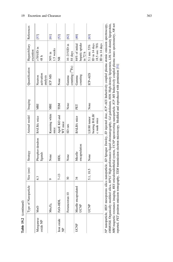
Table 19.2
(continued)
Type of Nanoparticle
Size (nm)
Strategy
Animal model
Imaging
Quantification
Hepatobiliary
excretion
References
Manganese
oxide NP
MnO
6.3
Phosphor-dendron
ligands
BALB/c mice
MRI
Neutron
activation
analysis
>70%ID in
48 h
[57]
Mn
3 O
4
9
None
Kunming white
mice
MRI
ICP–MS
50% in
1.5 weeks
[61]
Iron oxide
NP
FeO–HDL
7–13
HDL
ApoE KO and
WT mice
TEM
None
NR
[52]
Ferumoxtran-10
30
None
SD rats
None
Gamma
counting (
59
Fe)
16–21%ID in
84 days
[62]
UCNP
Micelle encapsulated
UCNP
34
Micelle
encapsulation
BALB/c mice
PET
Gamma
counting
84% of initial
hepatic uptake
in 72 h
[40]
UCNP
5.1, 18.5
None
LS180 tumor
bearing BALB/
c nude mice
None
ICP–AES
5.1 nm: 53%
ID in 14 days
18.5 nm: 78%
ID in 14 days
[63]
NP nanoparticle,
MSN mesoporous silica nanoparticle,
SD Sprague Dawley,
ID injected dose,
ICP–AES Inductively coupled plasma atomic emission spectroscopy,
ORMOSIL Organically modified silica,
HPLC High-performance liquid chromatography,
Gd gadolinium,
HDL High-density lipoprotein,
LDL low-density lipoprotein,
MRI magnetic resonance imaging,
RES reticuloendothelial system,
UCNP upconverting nanoparticle,
ICP–MS Inductively coupled plasma mass spectrometry,
NR not
reported,
PET positron emission tomography,
TEM transmission electron microscopy. Modified and reproduced with permission [51]
19 Excretion and Clearance
363
(continued)
Type of Nanoparticle
Size (nm)
Strategy
Animal model
Imaging
Quantification
Hepatobiliary
excretion
References
Manganese
oxide NP
MnO
6.3
Phosphor-dendron
ligands
BALB/c mice
MRI
Neutron
activation
analysis
>70%ID in
48 h
[57]
Mn
3 O
4
9
None
Kunming white
mice
MRI
ICP–MS
50% in
1.5 weeks
[61]
Iron oxide
NP
FeO–HDL
7–13
HDL
ApoE KO and
WT mice
TEM
None
NR
[52]
Ferumoxtran-10
30
None
SD rats
None
Gamma
counting (
59
Fe)
16–21%ID in
84 days
[62]
UCNP
Micelle encapsulated
UCNP
34
Micelle
encapsulation
BALB/c mice
PET
Gamma
counting
84% of initial
hepatic uptake
in 72 h
[40]
UCNP
5.1, 18.5
None
LS180 tumor
bearing BALB/
c nude mice
None
ICP–AES
5.1 nm: 53%
ID in 14 days
18.5 nm: 78%
ID in 14 days
[63]
NP nanoparticle,
MSN mesoporous silica nanoparticle,
SD Sprague Dawley,
ID injected dose,
ICP–AES Inductively coupled plasma atomic emission spectroscopy,
ORMOSIL Organically modified silica,
HPLC High-performance liquid chromatography,
Gd gadolinium,
HDL High-density lipoprotein,
LDL low-density lipoprotein,
MRI magnetic resonance imaging,
RES reticuloendothelial system,
UCNP upconverting nanoparticle,
ICP–MS Inductively coupled plasma mass spectrometry,
NR not
reported,
PET positron emission tomography,
TEM transmission electron microscopy. Modified and reproduced with permission [51]
19 Excretion and Clearance
363