

If the NPs are not recognized by MPS and have a smaller size than 120 nm (size
of sinusoidal fenestration), there is a high chance for NPs to be taken up by
hepatocytes and excreted via a hepatobiliary pathway. Thus, the strategy to enhance
the hepatobiliary excretion of the NPs is similar to that of lowering MPS recognition. Surface modification such as higher charge or micelle encapsulation can
improve the efficiency of hepatobiliary excretion. Souris et al. reported that
mesoporous silica NPs with higher positive surface charge (+34.4 mV) showed
faster hepatobiliary excretion than those with negative surface charge (−17.6 mV)
[53]. In vivo biodistribution analysis, substantial excretion to intestine was
observed after initial uptake to the liver [40, 53]. Seo et al. reported the micelle
encapsulated upconverting NP (UCNP) can be cleared via hepatobiliary excretion
in their integral forms [40]. We observed the substantial amount of NPs were
excreted from liver after initial liver accumulation. It is speculated that micelle
encapsulation of the NPs may lower the protein corona formation and thus lower
MPS recognition.
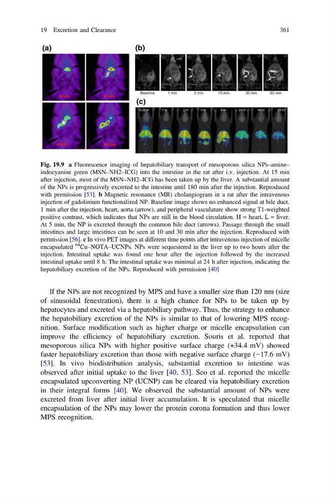
Fig. 19.9 a Fluorescence imaging of hepatobiliary transport of mesoporous silica NPs–amine–
indocyanine green (MSN–NH2–ICG) into the intestine in the rat after i.v. injection. At 15 min
after injection, most of the MSN–NH2–ICG has been taken up by the liver. A substantial amount
of the NPs is progressively excreted to the intestine until 180 min after the injection. Reproduced
with permission [53]. b Magnetic resonance (MR) cholangiogram in a rat after the intravenous
injection of gadolinium functionalized NP. Baseline image shows no enhanced signal at bile duct.
1 min after the injection, heart, aorta (arrow), and peripheral vasculature show strong T1-weighted
positive contrast, which indicates that NPs are still in the blood circulation. H = heart, L = liver.
At 5 min, the NP is excreted through the common bile duct (arrows). Passage through the small
intestines and large intestines can be seen at 10 and 30 min after the injection. Reproduced with
permission [56]. c In vivo PET images at different time points after intravenous injection of micelle
encapsulated
64
Cu–NOTA–UCNPs. NPs were sequestered in the liver up to two hours after the
injection. Intestinal uptake was found one hour after the injection followed by the increased
intestinal uptake until 8 h. The intestinal uptake was minimal at 24 h after injection, indicating the
hepatobiliary excretion of the NPs. Reproduced with permission [40]
19 Excretion and Clearance
361
of sinusoidal fenestration), there is a high chance for NPs to be taken up by
hepatocytes and excreted via a hepatobiliary pathway. Thus, the strategy to enhance
the hepatobiliary excretion of the NPs is similar to that of lowering MPS recognition. Surface modification such as higher charge or micelle encapsulation can
improve the efficiency of hepatobiliary excretion. Souris et al. reported that
mesoporous silica NPs with higher positive surface charge (+34.4 mV) showed
faster hepatobiliary excretion than those with negative surface charge (−17.6 mV)
[53]. In vivo biodistribution analysis, substantial excretion to intestine was
observed after initial uptake to the liver [40, 53]. Seo et al. reported the micelle
encapsulated upconverting NP (UCNP) can be cleared via hepatobiliary excretion
in their integral forms [40]. We observed the substantial amount of NPs were
excreted from liver after initial liver accumulation. It is speculated that micelle
encapsulation of the NPs may lower the protein corona formation and thus lower
MPS recognition.
Fig. 19.9 a Fluorescence imaging of hepatobiliary transport of mesoporous silica NPs–amine–
indocyanine green (MSN–NH2–ICG) into the intestine in the rat after i.v. injection. At 15 min
after injection, most of the MSN–NH2–ICG has been taken up by the liver. A substantial amount
of the NPs is progressively excreted to the intestine until 180 min after the injection. Reproduced
with permission [53]. b Magnetic resonance (MR) cholangiogram in a rat after the intravenous
injection of gadolinium functionalized NP. Baseline image shows no enhanced signal at bile duct.
1 min after the injection, heart, aorta (arrow), and peripheral vasculature show strong T1-weighted
positive contrast, which indicates that NPs are still in the blood circulation. H = heart, L = liver.
At 5 min, the NP is excreted through the common bile duct (arrows). Passage through the small
intestines and large intestines can be seen at 10 and 30 min after the injection. Reproduced with
permission [56]. c In vivo PET images at different time points after intravenous injection of micelle
encapsulated
64
Cu–NOTA–UCNPs. NPs were sequestered in the liver up to two hours after the
injection. Intestinal uptake was found one hour after the injection followed by the increased
intestinal uptake until 8 h. The intestinal uptake was minimal at 24 h after injection, indicating the
hepatobiliary excretion of the NPs. Reproduced with permission [40]
19 Excretion and Clearance
361