

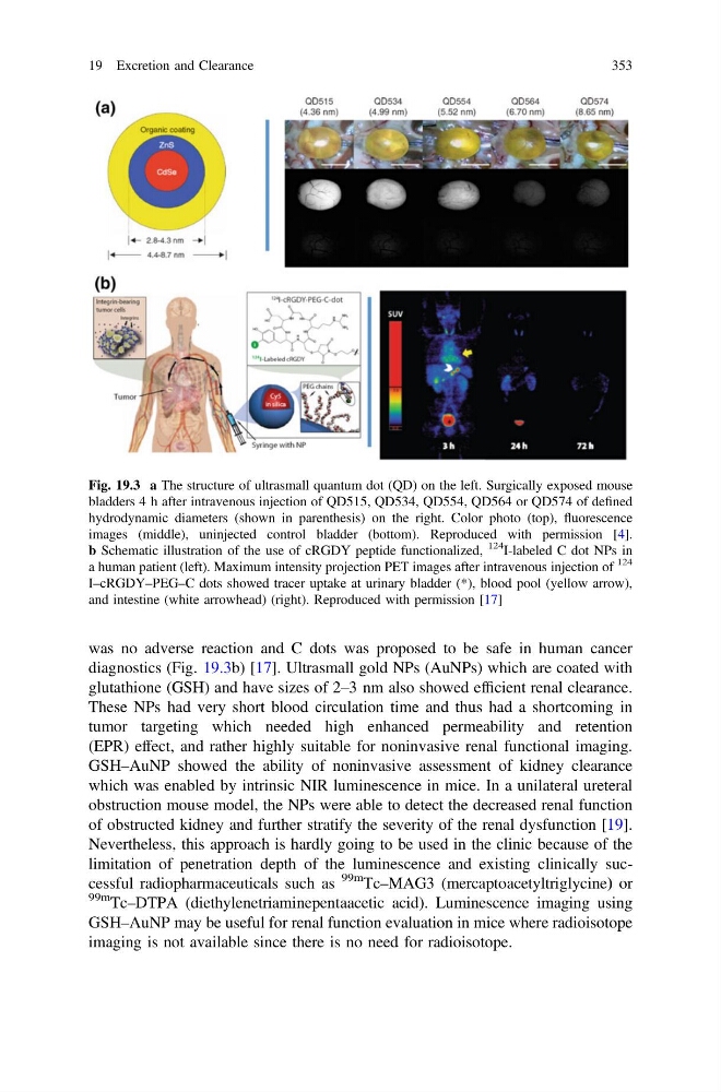
was no adverse reaction and C dots was proposed to be safe in human cancer
diagnostics (Fig. 19.3b) [17]. Ultrasmall gold NPs (AuNPs) which are coated with
glutathione (GSH) and have sizes of 2–3 nm also showed efficient renal clearance.
These NPs had very short blood circulation time and thus had a shortcoming in
tumor targeting which needed high enhanced permeability and retention
(EPR) effect, and rather highly suitable for noninvasive renal functional imaging.
GSH–AuNP showed the ability of noninvasive assessment of kidney clearance
which was enabled by intrinsic NIR luminescence in mice. In a unilateral ureteral
obstruction mouse model, the NPs were able to detect the decreased renal function
of obstructed kidney and further stratify the severity of the renal dysfunction [19].
Nevertheless, this approach is hardly going to be used in the clinic because of the
limitation of penetration depth of the luminescence and existing clinically successful radiopharmaceuticals such as
99m
Tc–MAG3 (mercaptoacetyltriglycine) or
99m Tc–DTPA (diethylenetriaminepentaacetic acid). Luminescence imaging using
GSH–AuNP may be useful for renal function evaluation in mice where radioisotope
imaging is not available since there is no need for radioisotope.
Fig. 19.3 a The structure of ultrasmall quantum dot (QD) on the left. Surgically exposed mouse
bladders 4 h after intravenous injection of QD515, QD534, QD554, QD564 or QD574 of defined
hydrodynamic diameters (shown in parenthesis) on the right. Color photo (top), fluorescence
images (middle), uninjected control bladder (bottom). Reproduced with permission [4].
b Schematic illustration of the use of cRGDY peptide functionalized,
124
I-labeled C dot NPs in
a human patient (left). Maximum intensity projection PET images after intravenous injection of
124
I–cRGDY–PEG–C dots showed tracer uptake at urinary bladder (*), blood pool (yellow arrow),
and intestine (white arrowhead) (right). Reproduced with permission [17]
19 Excretion and Clearance
353
diagnostics (Fig. 19.3b) [17]. Ultrasmall gold NPs (AuNPs) which are coated with
glutathione (GSH) and have sizes of 2–3 nm also showed efficient renal clearance.
These NPs had very short blood circulation time and thus had a shortcoming in
tumor targeting which needed high enhanced permeability and retention
(EPR) effect, and rather highly suitable for noninvasive renal functional imaging.
GSH–AuNP showed the ability of noninvasive assessment of kidney clearance
which was enabled by intrinsic NIR luminescence in mice. In a unilateral ureteral
obstruction mouse model, the NPs were able to detect the decreased renal function
of obstructed kidney and further stratify the severity of the renal dysfunction [19].
Nevertheless, this approach is hardly going to be used in the clinic because of the
limitation of penetration depth of the luminescence and existing clinically successful radiopharmaceuticals such as
99m
Tc–MAG3 (mercaptoacetyltriglycine) or
99m Tc–DTPA (diethylenetriaminepentaacetic acid). Luminescence imaging using
GSH–AuNP may be useful for renal function evaluation in mice where radioisotope
imaging is not available since there is no need for radioisotope.
Fig. 19.3 a The structure of ultrasmall quantum dot (QD) on the left. Surgically exposed mouse
bladders 4 h after intravenous injection of QD515, QD534, QD554, QD564 or QD574 of defined
hydrodynamic diameters (shown in parenthesis) on the right. Color photo (top), fluorescence
images (middle), uninjected control bladder (bottom). Reproduced with permission [4].
b Schematic illustration of the use of cRGDY peptide functionalized,
124
I-labeled C dot NPs in
a human patient (left). Maximum intensity projection PET images after intravenous injection of
124
I–cRGDY–PEG–C dots showed tracer uptake at urinary bladder (*), blood pool (yellow arrow),
and intestine (white arrowhead) (right). Reproduced with permission [17]
19 Excretion and Clearance
353