

active targeting of the tumor by attaching tumor-specific ligands to nanoparticles
enhances the nanoparticle potency [1, 51–53]. Barlett et al. comparatively evaluated
the in vivo biodistribution and functional activity of siRNA targeting (silencing)
luciferase mRNA delivered by transferrin-targeted or non-targeted
DOTA-conjugated nanoparticles labeled with
64 Cu [54]. The results of the PET/
CT study demonstrate the negligible impact of transferrin targeting on the accumulation of nanoparticles in the luciferase-expressing Neuro2A s.c. tumors in mice.
However, the active targeting altered the functional activity of the tumors (approximately 50% lower relative increase of tumor luciferase activity measured by
bioluminescent imaging in mice treated with transferrin targeted nanoparticles
compared to those treated with non-targeted nanoparticles). In the compartment
model analysis performed to explicate the discrepancy between biodistribution and
functional activity of tumor-specific targeting nanoparticles, the tumor tissue time–
activity curves were reasonably fitted with zero k 2 . Also, one-tissue and two-tissue
compartment models yielded equivalent curve-fitting qualities. In this situation,
where the radiotracers do not return to the bloodstream and C 1 (tumor interstitial
space in this specific model) and C 2 (specific tumor targeting) quickly attain the
equilibrium state (Fig. 16.1b), temporal changes in the total tissue activity measurable by PET is not influenced by changes in k 3 associated with the tumor-specific
binding. In addition, mathematical solutions of C 1 and C 2 composed with different
k 3 values and other fixed parameters enable us to comprehend how tumor-specific
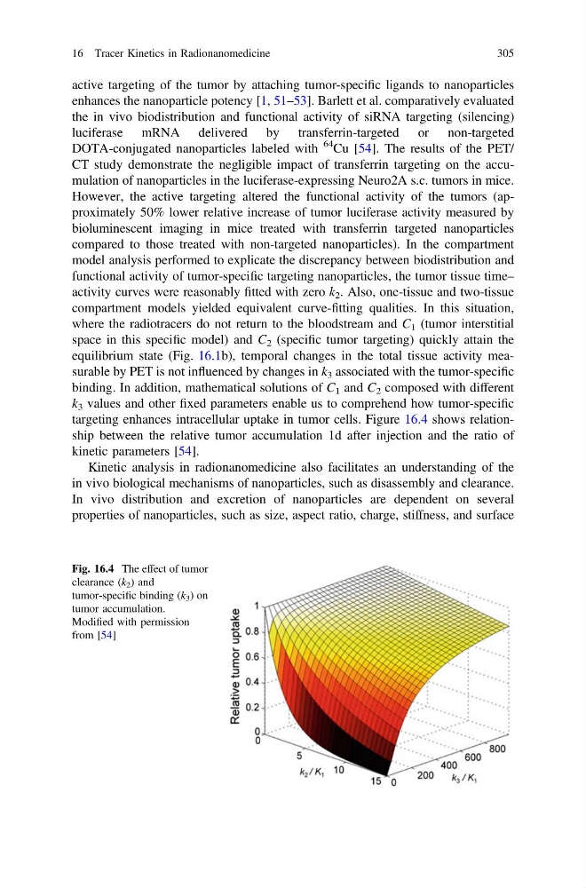
targeting enhances intracellular uptake in tumor cells. Figure 16.4 shows relationship between the relative tumor accumulation 1d after injection and the ratio of
kinetic parameters [54].
Kinetic analysis in radionanomedicine also facilitates an understanding of the
in vivo biological mechanisms of nanoparticles, such as disassembly and clearance.
In vivo distribution and excretion of nanoparticles are dependent on several
properties of nanoparticles, such as size, aspect ratio, charge, stiffness, and surface
Fig. 16.4 The effect of tumor
clearance (k 2 ) and
tumor-specific binding (k 3 ) on
tumor accumulation.
Modified with permission
from [54]
16 Tracer Kinetics in Radionanomedicine
305
enhances the nanoparticle potency [1, 51–53]. Barlett et al. comparatively evaluated
the in vivo biodistribution and functional activity of siRNA targeting (silencing)
luciferase mRNA delivered by transferrin-targeted or non-targeted
DOTA-conjugated nanoparticles labeled with
64 Cu [54]. The results of the PET/
CT study demonstrate the negligible impact of transferrin targeting on the accumulation of nanoparticles in the luciferase-expressing Neuro2A s.c. tumors in mice.
However, the active targeting altered the functional activity of the tumors (approximately 50% lower relative increase of tumor luciferase activity measured by
bioluminescent imaging in mice treated with transferrin targeted nanoparticles
compared to those treated with non-targeted nanoparticles). In the compartment
model analysis performed to explicate the discrepancy between biodistribution and
functional activity of tumor-specific targeting nanoparticles, the tumor tissue time–
activity curves were reasonably fitted with zero k 2 . Also, one-tissue and two-tissue
compartment models yielded equivalent curve-fitting qualities. In this situation,
where the radiotracers do not return to the bloodstream and C 1 (tumor interstitial
space in this specific model) and C 2 (specific tumor targeting) quickly attain the
equilibrium state (Fig. 16.1b), temporal changes in the total tissue activity measurable by PET is not influenced by changes in k 3 associated with the tumor-specific
binding. In addition, mathematical solutions of C 1 and C 2 composed with different
k 3 values and other fixed parameters enable us to comprehend how tumor-specific
targeting enhances intracellular uptake in tumor cells. Figure 16.4 shows relationship between the relative tumor accumulation 1d after injection and the ratio of
kinetic parameters [54].
Kinetic analysis in radionanomedicine also facilitates an understanding of the
in vivo biological mechanisms of nanoparticles, such as disassembly and clearance.
In vivo distribution and excretion of nanoparticles are dependent on several
properties of nanoparticles, such as size, aspect ratio, charge, stiffness, and surface
Fig. 16.4 The effect of tumor
clearance (k 2 ) and
tumor-specific binding (k 3 ) on
tumor accumulation.
Modified with permission
from [54]
16 Tracer Kinetics in Radionanomedicine
305