

chemistry [2]. Zuckerman et al. investigated the mechanism of the unexpected rapid
renal clearance of cationic cyclodextrin-containing polymer (CDP)-based siRNA
nanoparticles. This mechanism prevents nanoparticles from prolonging the siRNA
delivery to the tumor thereby enhancing the anticancer efficacy. These siRNA
nanoparticles were designed to have a much bigger size (D % 100 nm) than the
effective cut-off size of the renal filtration barrier (D % 10 nm) [55]. Microscopic
imaging studies conducted on mice have demonstrated that CDP-based siRNA
nanoparticles transiently accumulate and can be dissembled by the glomerular
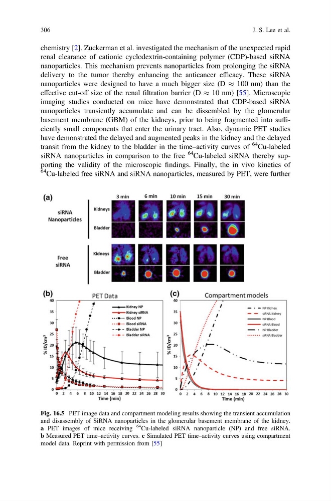
basement membrane (GBM) of the kidneys, prior to being fragmented into sufficiently small components that enter the urinary tract. Also, dynamic PET studies
have demonstrated the delayed and augmented peaks in the kidney and the delayed
transit from the kidney to the bladder in the time–activity curves of
64 Cu-labeled
siRNA nanoparticles in comparison to the free
64 Cu-labeled siRNA thereby supporting the validity of the microscopic findings. Finally, the in vivo kinetics of
64 Cu-labeled free siRNA and siRNA nanoparticles, measured by PET, were further
Fig. 16.5 PET image data and compartment modeling results showing the transient accumulation
and disassembly of SiRNA nanoparticles in the glomerular basement membrane of the kidney.
a PET images of mice receiving
64
Cu-labeled siRNA nanoparticle (NP) and free siRNA.
b Measured PET time–activity curves. c Simulated PET time–activity curves using compartment
model data. Reprint with permission from [55]
306
J. S. Lee et al.
renal clearance of cationic cyclodextrin-containing polymer (CDP)-based siRNA
nanoparticles. This mechanism prevents nanoparticles from prolonging the siRNA
delivery to the tumor thereby enhancing the anticancer efficacy. These siRNA
nanoparticles were designed to have a much bigger size (D % 100 nm) than the
effective cut-off size of the renal filtration barrier (D % 10 nm) [55]. Microscopic
imaging studies conducted on mice have demonstrated that CDP-based siRNA
nanoparticles transiently accumulate and can be dissembled by the glomerular
basement membrane (GBM) of the kidneys, prior to being fragmented into sufficiently small components that enter the urinary tract. Also, dynamic PET studies
have demonstrated the delayed and augmented peaks in the kidney and the delayed
transit from the kidney to the bladder in the time–activity curves of
64 Cu-labeled
siRNA nanoparticles in comparison to the free
64 Cu-labeled siRNA thereby supporting the validity of the microscopic findings. Finally, the in vivo kinetics of
64 Cu-labeled free siRNA and siRNA nanoparticles, measured by PET, were further
Fig. 16.5 PET image data and compartment modeling results showing the transient accumulation
and disassembly of SiRNA nanoparticles in the glomerular basement membrane of the kidney.
a PET images of mice receiving
64
Cu-labeled siRNA nanoparticle (NP) and free siRNA.
b Measured PET time–activity curves. c Simulated PET time–activity curves using compartment
model data. Reprint with permission from [55]
306
J. S. Lee et al.