

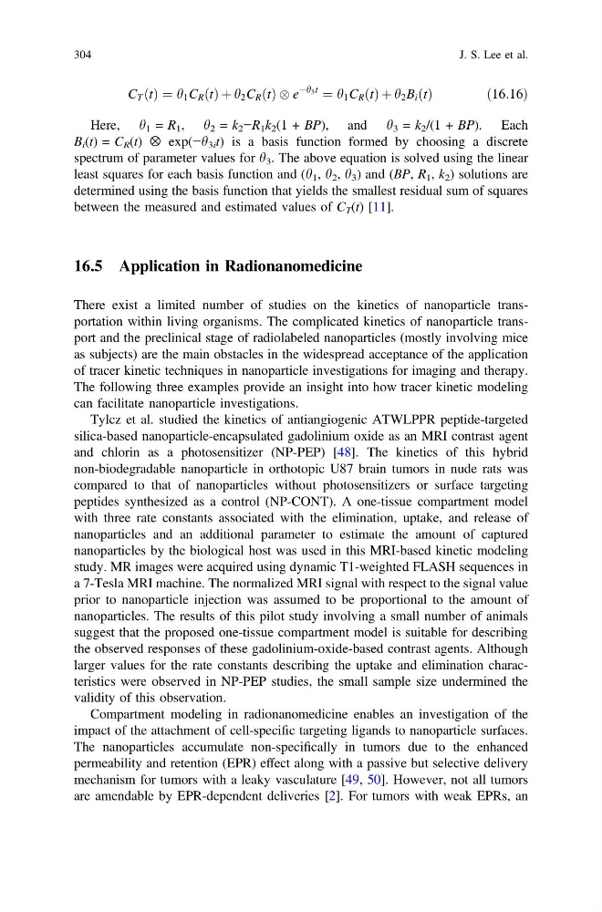
C T ðtÞ ¼ h 1 C R ðtÞ þ h 2 C R ðtÞ e
Àh 3 t
¼ h 1 C R ðtÞ þ h 2 B i ðtÞ
ð 16:16Þ
Here, h 1 = R 1 , h 2 = k 2 −R 1 k 2 (1 + BP), and h 3 = k 2 /(1 + BP). Each
B i (t) = C R (t) ⊗ exp(−h 3i t) is a basis function formed by choosing a discrete
spectrum of parameter values for h 3 . The above equation is solved using the linear
least squares for each basis function and (h 1 , h 2 , h 3 ) and (BP, R 1 , k 2 ) solutions are
determined using the basis function that yields the smallest residual sum of squares
between the measured and estimated values of C T (t) [11].
16.5 Application in Radionanomedicine
There exist a limited number of studies on the kinetics of nanoparticle transportation within living organisms. The complicated kinetics of nanoparticle transport and the preclinical stage of radiolabeled nanoparticles (mostly involving mice
as subjects) are the main obstacles in the widespread acceptance of the application
of tracer kinetic techniques in nanoparticle investigations for imaging and therapy.
The following three examples provide an insight into how tracer kinetic modeling
can facilitate nanoparticle investigations.
Tylcz et al. studied the kinetics of antiangiogenic ATWLPPR peptide-targeted
silica-based nanoparticle-encapsulated gadolinium oxide as an MRI contrast agent
and chlorin as a photosensitizer (NP-PEP) [48]. The kinetics of this hybrid
non-biodegradable nanoparticle in orthotopic U87 brain tumors in nude rats was
compared to that of nanoparticles without photosensitizers or surface targeting
peptides synthesized as a control (NP-CONT). A one-tissue compartment model
with three rate constants associated with the elimination, uptake, and release of
nanoparticles and an additional parameter to estimate the amount of captured
nanoparticles by the biological host was used in this MRI-based kinetic modeling
study. MR images were acquired using dynamic T1-weighted FLASH sequences in
a 7-Tesla MRI machine. The normalized MRI signal with respect to the signal value
prior to nanoparticle injection was assumed to be proportional to the amount of
nanoparticles. The results of this pilot study involving a small number of animals
suggest that the proposed one-tissue compartment model is suitable for describing
the observed responses of these gadolinium-oxide-based contrast agents. Although
larger values for the rate constants describing the uptake and elimination characteristics were observed in NP-PEP studies, the small sample size undermined the
validity of this observation.
Compartment modeling in radionanomedicine enables an investigation of the
impact of the attachment of cell-specific targeting ligands to nanoparticle surfaces.
The nanoparticles accumulate non-specifically in tumors due to the enhanced
permeability and retention (EPR) effect along with a passive but selective delivery
mechanism for tumors with a leaky vasculature [49, 50]. However, not all tumors
are amendable by EPR-dependent deliveries [2]. For tumors with weak EPRs, an
304
J. S. Lee et al.
Àh 3 t
¼ h 1 C R ðtÞ þ h 2 B i ðtÞ
ð 16:16Þ
Here, h 1 = R 1 , h 2 = k 2 −R 1 k 2 (1 + BP), and h 3 = k 2 /(1 + BP). Each
B i (t) = C R (t) ⊗ exp(−h 3i t) is a basis function formed by choosing a discrete
spectrum of parameter values for h 3 . The above equation is solved using the linear
least squares for each basis function and (h 1 , h 2 , h 3 ) and (BP, R 1 , k 2 ) solutions are
determined using the basis function that yields the smallest residual sum of squares
between the measured and estimated values of C T (t) [11].
16.5 Application in Radionanomedicine
There exist a limited number of studies on the kinetics of nanoparticle transportation within living organisms. The complicated kinetics of nanoparticle transport and the preclinical stage of radiolabeled nanoparticles (mostly involving mice
as subjects) are the main obstacles in the widespread acceptance of the application
of tracer kinetic techniques in nanoparticle investigations for imaging and therapy.
The following three examples provide an insight into how tracer kinetic modeling
can facilitate nanoparticle investigations.
Tylcz et al. studied the kinetics of antiangiogenic ATWLPPR peptide-targeted
silica-based nanoparticle-encapsulated gadolinium oxide as an MRI contrast agent
and chlorin as a photosensitizer (NP-PEP) [48]. The kinetics of this hybrid
non-biodegradable nanoparticle in orthotopic U87 brain tumors in nude rats was
compared to that of nanoparticles without photosensitizers or surface targeting
peptides synthesized as a control (NP-CONT). A one-tissue compartment model
with three rate constants associated with the elimination, uptake, and release of
nanoparticles and an additional parameter to estimate the amount of captured
nanoparticles by the biological host was used in this MRI-based kinetic modeling
study. MR images were acquired using dynamic T1-weighted FLASH sequences in
a 7-Tesla MRI machine. The normalized MRI signal with respect to the signal value
prior to nanoparticle injection was assumed to be proportional to the amount of
nanoparticles. The results of this pilot study involving a small number of animals
suggest that the proposed one-tissue compartment model is suitable for describing
the observed responses of these gadolinium-oxide-based contrast agents. Although
larger values for the rate constants describing the uptake and elimination characteristics were observed in NP-PEP studies, the small sample size undermined the
validity of this observation.
Compartment modeling in radionanomedicine enables an investigation of the
impact of the attachment of cell-specific targeting ligands to nanoparticle surfaces.
The nanoparticles accumulate non-specifically in tumors due to the enhanced
permeability and retention (EPR) effect along with a passive but selective delivery
mechanism for tumors with a leaky vasculature [49, 50]. However, not all tumors
are amendable by EPR-dependent deliveries [2]. For tumors with weak EPRs, an
304
J. S. Lee et al.