

improvement, minimal amount of the NPs (0.02%ID/g) was able to enter inside of the
brain [59]. BBB is destroyed in the brain cancer, thus targeting NPs to brain cancer is
more feasible than normal brain tissue. Seo et al. reported that
64 Cu labeled liposome
and micelle can be targeted to brain cancer lesion in orthotopic glioblastoma model of
mouse. They also found that 20 nm sized micelles showed the higher uptake than
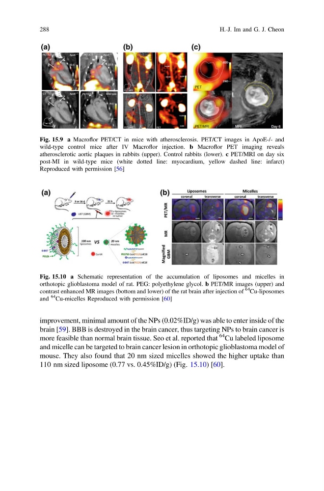
110 nm sized liposome (0.77 vs. 0.45%ID/g) (Fig. 15.10) [60].
Fig. 15.9 a Macroflor PET/CT in mice with atherosclerosis. PET/CT images in ApoE-/- and
wild-type control mice after IV Macroflor injection. b Macroflor PET imaging reveals
atherosclerotic aortic plaques in rabbits (upper). Control rabbits (lower). c PET/MRI on day six
post-MI in wild-type mice (white dotted line: myocardium, yellow dashed line: infarct)
Reproduced with permission [56]
Fig. 15.10 a Schematic representation of the accumulation of liposomes and micelles in
orthotopic glioblastoma model of rat. PEG: polyethylene glycol. b PET/MR images (upper) and
contrast enhanced MR images (bottom and lower) of the rat brain after injection of
64
Cu-liposomes
and
64
Cu-micelles Reproduced with permission [60]
288
H.-J. Im and G. J. Cheon
brain [59]. BBB is destroyed in the brain cancer, thus targeting NPs to brain cancer is
more feasible than normal brain tissue. Seo et al. reported that
64 Cu labeled liposome
and micelle can be targeted to brain cancer lesion in orthotopic glioblastoma model of
mouse. They also found that 20 nm sized micelles showed the higher uptake than
110 nm sized liposome (0.77 vs. 0.45%ID/g) (Fig. 15.10) [60].
Fig. 15.9 a Macroflor PET/CT in mice with atherosclerosis. PET/CT images in ApoE-/- and
wild-type control mice after IV Macroflor injection. b Macroflor PET imaging reveals
atherosclerotic aortic plaques in rabbits (upper). Control rabbits (lower). c PET/MRI on day six
post-MI in wild-type mice (white dotted line: myocardium, yellow dashed line: infarct)
Reproduced with permission [56]
Fig. 15.10 a Schematic representation of the accumulation of liposomes and micelles in
orthotopic glioblastoma model of rat. PEG: polyethylene glycol. b PET/MR images (upper) and
contrast enhanced MR images (bottom and lower) of the rat brain after injection of
64
Cu-liposomes
and
64
Cu-micelles Reproduced with permission [60]
288
H.-J. Im and G. J. Cheon