

15.3.2 PET/CT, PET/MR Imaging in Radionanomedicine
PET has also been utilized in radionanomedicine for quantitative evaluation of
in vivo biodistribution including targeting efficiency. Majority of the studies were
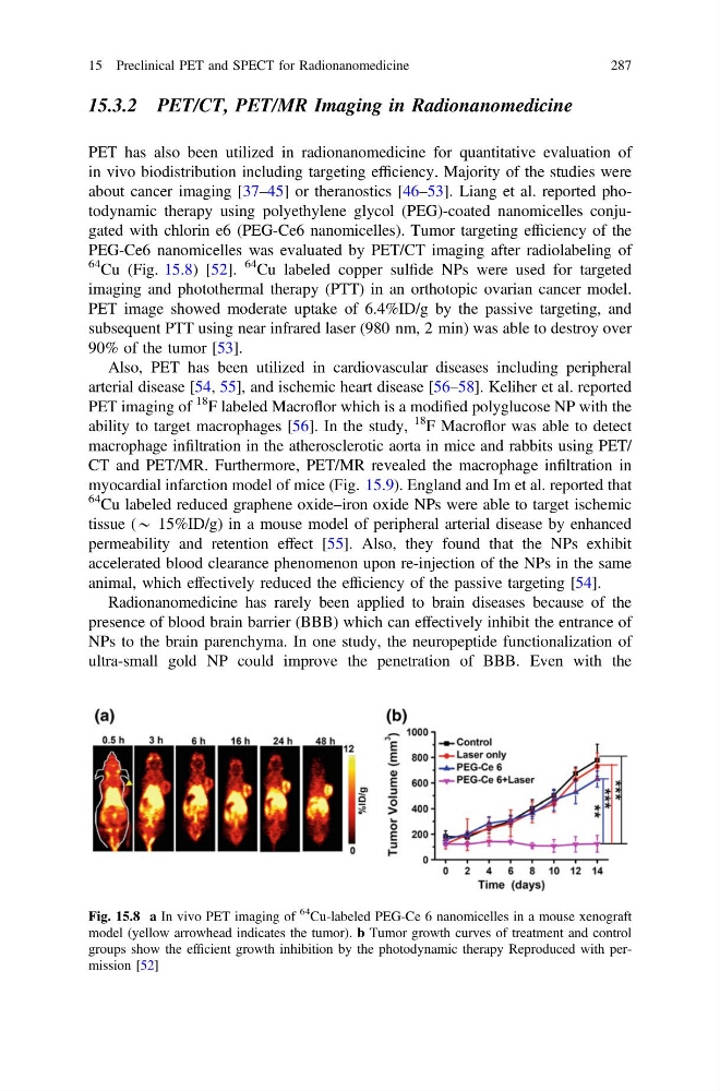
about cancer imaging [37–45] or theranostics [46–53]. Liang et al. reported photodynamic therapy using polyethylene glycol (PEG)-coated nanomicelles conjugated with chlorin e6 (PEG-Ce6 nanomicelles). Tumor targeting efficiency of the
PEG-Ce6 nanomicelles was evaluated by PET/CT imaging after radiolabeling of
64 Cu (Fig. 15.8) [52].
64 Cu labeled copper sulfide NPs were used for targeted
imaging and photothermal therapy (PTT) in an orthotopic ovarian cancer model.
PET image showed moderate uptake of 6.4%ID/g by the passive targeting, and
subsequent PTT using near infrared laser (980 nm, 2 min) was able to destroy over
90% of the tumor [53].
Also, PET has been utilized in cardiovascular diseases including peripheral
arterial disease [54, 55], and ischemic heart disease [56–58]. Keliher et al. reported
PET imaging of
18 F labeled Macroflor which is a modified polyglucose NP with the
ability to target macrophages [56]. In the study,
18 F Macroflor was able to detect
macrophage infiltration in the atherosclerotic aorta in mice and rabbits using PET/
CT and PET/MR. Furthermore, PET/MR revealed the macrophage infiltration in
myocardial infarction model of mice (Fig. 15.9). England and Im et al. reported that
64 Cu labeled reduced graphene oxide–iron oxide NPs were able to target ischemic
tissue (* 15%ID/g) in a mouse model of peripheral arterial disease by enhanced
permeability and retention effect [55]. Also, they found that the NPs exhibit
accelerated blood clearance phenomenon upon re-injection of the NPs in the same
animal, which effectively reduced the efficiency of the passive targeting [54].
Radionanomedicine has rarely been applied to brain diseases because of the
presence of blood brain barrier (BBB) which can effectively inhibit the entrance of
NPs to the brain parenchyma. In one study, the neuropeptide functionalization of
ultra-small gold NP could improve the penetration of BBB. Even with the
Fig. 15.8 a In vivo PET imaging of
64
Cu-labeled PEG-Ce 6 nanomicelles in a mouse xenograft
model (yellow arrowhead indicates the tumor). b Tumor growth curves of treatment and control
groups show the efficient growth inhibition by the photodynamic therapy Reproduced with permission [52]
15 Preclinical PET and SPECT for Radionanomedicine
287
PET has also been utilized in radionanomedicine for quantitative evaluation of
in vivo biodistribution including targeting efficiency. Majority of the studies were
about cancer imaging [37–45] or theranostics [46–53]. Liang et al. reported photodynamic therapy using polyethylene glycol (PEG)-coated nanomicelles conjugated with chlorin e6 (PEG-Ce6 nanomicelles). Tumor targeting efficiency of the
PEG-Ce6 nanomicelles was evaluated by PET/CT imaging after radiolabeling of
64 Cu (Fig. 15.8) [52].
64 Cu labeled copper sulfide NPs were used for targeted
imaging and photothermal therapy (PTT) in an orthotopic ovarian cancer model.
PET image showed moderate uptake of 6.4%ID/g by the passive targeting, and
subsequent PTT using near infrared laser (980 nm, 2 min) was able to destroy over
90% of the tumor [53].
Also, PET has been utilized in cardiovascular diseases including peripheral
arterial disease [54, 55], and ischemic heart disease [56–58]. Keliher et al. reported
PET imaging of
18 F labeled Macroflor which is a modified polyglucose NP with the
ability to target macrophages [56]. In the study,
18 F Macroflor was able to detect
macrophage infiltration in the atherosclerotic aorta in mice and rabbits using PET/
CT and PET/MR. Furthermore, PET/MR revealed the macrophage infiltration in
myocardial infarction model of mice (Fig. 15.9). England and Im et al. reported that
64 Cu labeled reduced graphene oxide–iron oxide NPs were able to target ischemic
tissue (* 15%ID/g) in a mouse model of peripheral arterial disease by enhanced
permeability and retention effect [55]. Also, they found that the NPs exhibit
accelerated blood clearance phenomenon upon re-injection of the NPs in the same
animal, which effectively reduced the efficiency of the passive targeting [54].
Radionanomedicine has rarely been applied to brain diseases because of the
presence of blood brain barrier (BBB) which can effectively inhibit the entrance of
NPs to the brain parenchyma. In one study, the neuropeptide functionalization of
ultra-small gold NP could improve the penetration of BBB. Even with the
Fig. 15.8 a In vivo PET imaging of
64
Cu-labeled PEG-Ce 6 nanomicelles in a mouse xenograft
model (yellow arrowhead indicates the tumor). b Tumor growth curves of treatment and control
groups show the efficient growth inhibition by the photodynamic therapy Reproduced with permission [52]
15 Preclinical PET and SPECT for Radionanomedicine
287